. 2018 Nov 3;20(1):247.
doi: 10.1186/s13075-018-1744-3.

The involvement of C5a in the progression of experimental arthritis with Porphyromonas gingivalis infection in SKG mice

Affiliations

The involvement of C5a in the progression of experimental arthritis with Porphyromonas gingivalis infection in SKG mice

Syuichi Munenaga et al. Arthritis Res Ther. .

Abstract

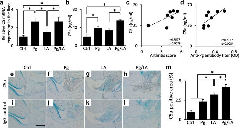
Background: Epidemiological evidence to suggest that periodontal disease (PD) is involved in the progression of rheumatoid arthritis (RA) is increasing. The complement system plays a critical role in immune responses. C5a has been implicated in chronic inflammatory diseases, including PD and RA. Porphyromonas gingivalis is the major causative bacteria of PD and can produce C5a. Therefore, it is hypothesized that P. gingivalis infection is involved in the progression of RA by elevating C5a levels. In the present study, P. gingivalis-infected RA model mice were established to investigate the involvement of C5a.

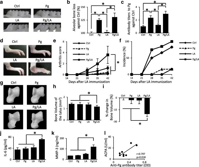
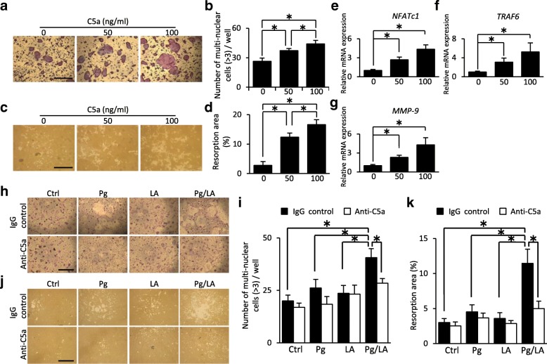
Methods: SKG mice orally infected with P. gingivalis were immunized with intraperitoneal injection of laminarin (LA) to induce arthritis. Arthritis development was assessed by arthritis score (AS), bone destruction on the talus, histology, and serum markers of RA. In order to investigate the effects of serum C5a on bone destruction, osteoclast differentiation of bone marrow mononuclear cells was examined by using serum samples from each group of mice. The relationship between C5a levels and antibody titers to periodontal pathogens in patients with RA was investigated by enzyme-linked immunosorbent assay.

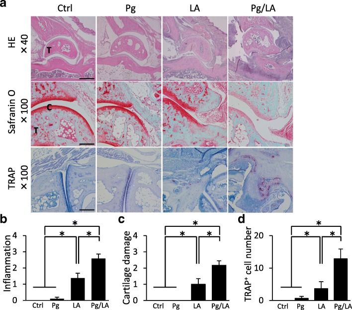
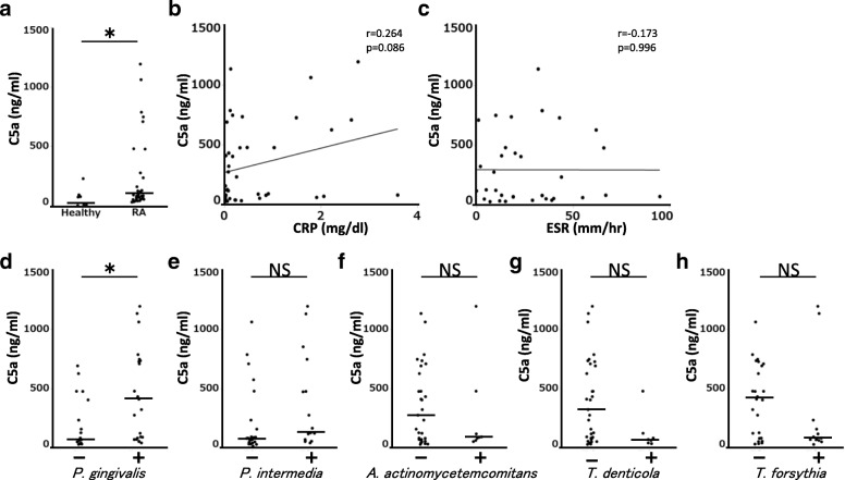
Results: P. gingivalis oral infection increased AS, infiltration of inflammatory cells, bone destruction on the talus, and serum markers of RA in mice immunized with LA. The addition of serum from LA-injected mice with the P. gingivalis oral infection promoted osteoclast differentiation, and the addition of a neutralization antibody against C5a suppressed osteoclast differentiation. C5a levels of serum in RA patients with positive P. gingivalis antibody were elevated compared with those in RA patients with negative P. gingivalis antibody.

Conclusions: These results suggest that P. gingivalis infection enhances the progression of RA via C5a.

Keywords: Arthritis; C5a; Porphyromonas gingivalis; SKG mice.


Conflict of interest statement

Ethics approval and consent to participate

Animal experiments were approved by the ethics committee of Hiroshima University (approval A12–15). All patients provided written informed consent prior to enrollment. This study was approved by the ethics committee of Hiroshima University Hospital (#1017).

Consent for publication

The consent of all coauthors was collected before submission.

Competing interests

The authors declare that they have no competing interests.

Publisher’s Note

Springer Nature remains neutral with regard to jurisdictional claims in published maps and institutional affiliations.

Figures

Fig. 1
Fig. 1
SKG mice received an oral inoculation of Porphyromonas gingivalis (Pg) (108 colony-forming units per mouse) repeatedly every 3 days for 42 days after the laminarin (LA) immunization: control (Ctrl) n = 6, Pg n = 6, LA n = 6, Pg/LA n = 8. a Representative images of the palatal surfaces of the maxillary molars from the four groups (arrows indicate the resorption site). b Alveolar bone loss was analyzed by measuring the distance from the cemento-enamel junction to the alveolar bone crest on palatal surfaces. c Antibody titers to P. gingivalis in serum were measured by enzyme-linked immunosorbent assay. d Representative figures of hind paws for the four groups showing joint inflammation. e, f The severity and incidence of arthritis were scored by using Sakaguchi’s arthritis score. g Representative micro-computed tomography images of the talus. h, i Bone volume of the talus and percentage changes in bone volume relative to Ctrl mice. j, k Serum concentrations of interleukin-6 (IL-6) and matrix metalloproteinase-3 (MMP-3) in the four groups. l The correlation between the serum level of anti-citrullinated protein antibody (ACPA) in the Pg/LA group and anti-Pg antibody titer. Data represent the mean ± standard deviation (b, c, and d) or mean ± standard error of the mean (a, and e–k) of 6–8 mice per group. Statistical analyses were performed by using the Tukey-Kramer test, Bonferroni-corrected Mann–Whitney U test, or Pearson’s correlation coefficient (*P <0.05). Abbreviation: OD optical density
Fig. 2
Fig. 2
Histological assessment of ankle joints on day 42 from each group of mice. a Representative sections of the ankle joint stained with hematoxylin and eosin (HE), Safranin O, and tartrate-resistant acid phosphatase (TRAP). b–d Histological scores of inflammation, cartilage damage, and TRAP-positive cell numbers from each group of mice (n = 6 per group). Data represent the mean ± standard error of the mean. Statistical analyses were performed by the Bonferroni-corrected Mann–Whitney U test (*P <0.05). Original magnifications: 40×; scale bar = 500 μm, 100×; scale bar = 250 μm. Abbreviations: C cartilage, Ctrl control, LA laminarin, Pg Porphyromonas gingivalis, T talus
Fig. 3
Fig. 3
Serum, liver, and joints were collected from each group of mice at the end point of the experiment. a The mRNA expression of C5 in the liver was measured by quantitative reverse transcription-polymerase chain reaction. b Serum concentrations of C5a were measured by enzyme-linked immunosorbent assay. c, d Relationship between C5a levels and arthritis score or anti–Porphyromonas gingivalis antibody titers in the Pg/LA group. e–l Representative immunohistochemical images of C5a in the ankle joint. m The C5a-positive area was measured by ImageJ (n = 6 per group). Data represent the mean ± standard error of the mean. Statistical analyses were performed by Tukey-Kramer test, Pearson’s correlation coefficient (d), and Spearman’s correlation coefficient (c) (*P <0.05). Abbreviations: Ctrl control, LA laminarin, OD optical density, Pg Porphyromonas gingivalis
Fig. 4
Fig. 4
Bone marrow mononuclear cells (BMCs) were cultured in the presence of macrophage colony-stimulating factor and soluble recombinant receptor activation of nuclear factor kappa-B ligand to promote osteoclast differentiation. a Representative images of osteoclast differentiation with recombinant C5a (rC5a) by tartrate-resistant acid phosphatase staining. b The number of multi-nuclear osteoclast in each well (n = 10). c Representative images of bone resorption in the presence of rC5a by pit formation assay. d Bone resorption area by multi-nuclear osteoclast in each well was measured by ImageJ (n = 5). e–g Quantitative reverse transcription-polymerase chain reaction analysis of NFATc1, TRAF6, and MMP-9 (n = 8). h Representative images of osteoclast differentiation of BMCs by the effect of serum derived from each group of mice in the presence of an isotype control antibody or anti-C5a antibody. i The number of multi-nuclear osteoclast in each well with or without serum derived from each group of mice (n = 10). j Representative images of bone resorption by the effect of serum derived from each group of mice in the presence of an isotype control antibody or anti-C5a antibody. k Bone resorption area by multi-nuclear osteoclast in each well was measured by ImageJ (n = 5). Statistical analyses were performed by the Student’s unpaired t test or Tukey-Kramer test (*P <0.05). Data represent the mean ± standard error of the mean. Original magnification: 40×; scale bar = 1000 μm
Fig. 5
Fig. 5
a C5a levels in serum from patients with rheumatoid arthritis (RA) and healthy control patients were measured by enzyme-linked immunosorbent assay (ELISA) (healthy, n = 8; patients with RA, n = 40). b, c Relationship between C5a levels and C-reactive protein (CRP) or erythrocyte sedimentation rate (ESR) in the serum from patients with RA. d–h C5a levels and antibody titers to periodontal pathogens (Porphyromonas gingivalis W83, Prevotella intermedia, Treponema denticola, Tannerella forsythia, and Aggregatibactor actinomyvetemcomitance) in serum from patients with RA were measured by ELISA. Bars show median of the data. Statistical analyses were performed by using Mann–Whitney U test and Pearson’s correlation coefficient (*P <0.05). Abbreviation: NS not significant

Similar articles

Cited by

References

    1. McInnes IB, Schett G. The pathogenesis of rheumatoid arthritis. N Engl J Med. 2011;365:2205–2219. doi: 10.1056/NEJMra1004965. - DOI - PubMed
    1. Goronzy JJ, Weyand CM. Developments in the scientific understanding of rheumatoid arthritis. Arthritis Res Ther. 2009;11:249. doi: 10.1186/ar2758. - DOI - PMC - PubMed
    1. Socransky SS, Haffajee AD. Implications of periodontal microbiology for the treatment of periodontal infections. Compend Suppl. 1994:S684–5 688–693; quiz S714–687. - PubMed
    1. Han YW, Wang X. Mobile microbiome: oral bacteria in extra-oral infections and inflammation. J Dent Res. 2013;92:485–491. doi: 10.1177/0022034513487559. - DOI - PMC - PubMed
    1. Scher JU, Bretz WA, Abramson SB. Periodontal disease and subgingival microbiota as contributors for rheumatoid arthritis pathogenesis: modifiable risk factors? Curr Opin Rheumatol. 2014;26:424–429. doi: 10.1097/BOR.0000000000000076. - DOI - PMC - PubMed

Publication types