

Nerve ultrasound characterizes AMN polyneuropathy as inhomogeneous and focal hypertrophic
- PMID: 30390710
- PMCID: PMC6215661
- DOI: 10.1186/s13023-018-0939-7
Nerve ultrasound characterizes AMN polyneuropathy as inhomogeneous and focal hypertrophic
Abstract
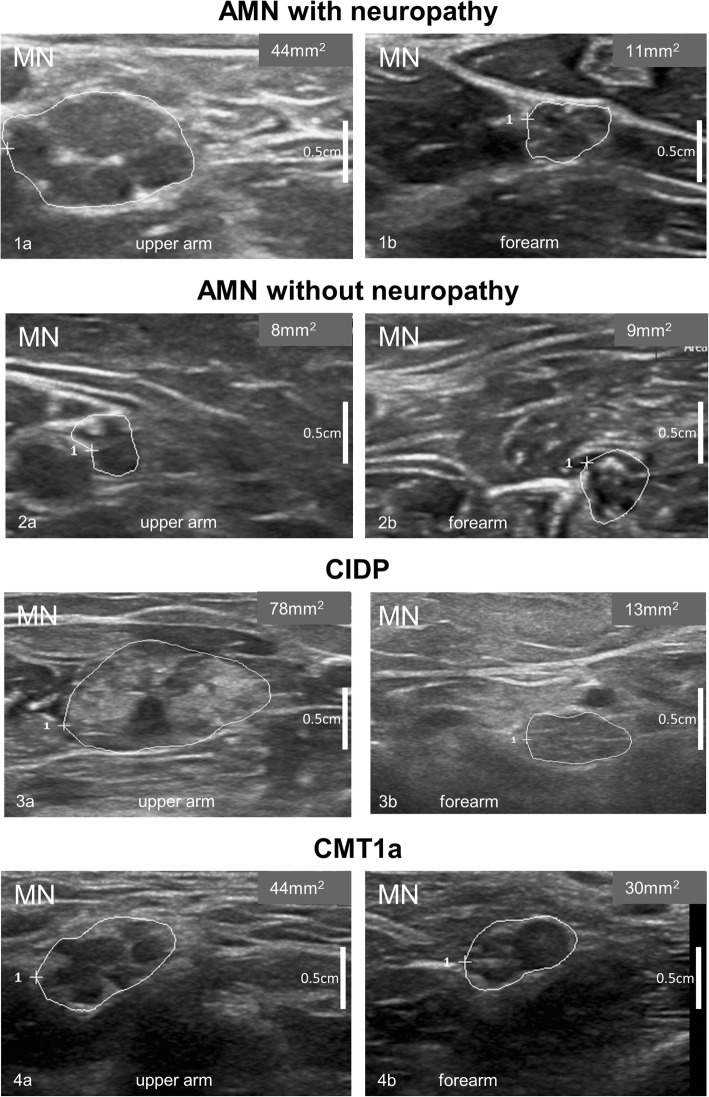
Objective: High-resolution nerve ultrasound (HRUS) is a painless tool to quickly evaluate peripheral nerve morphology in vivo. This study set out to characterize peripheral nerve involvement in X-linked adrenomyeloneuropathy (AMN) by HRUS.
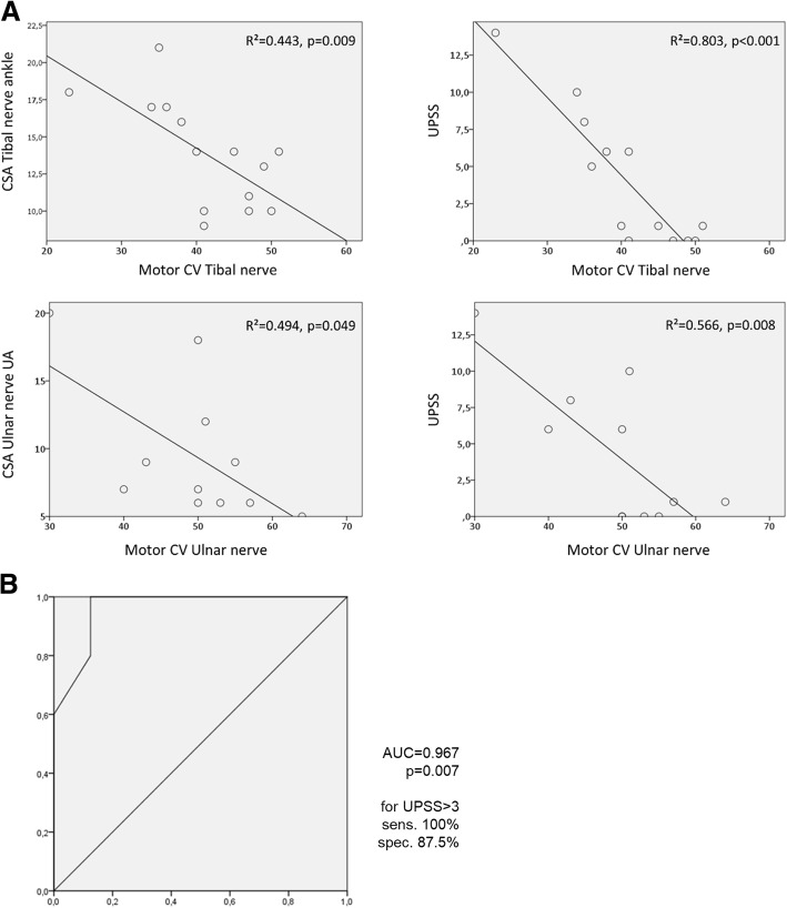
Methods: Thirteen adults with genetically proven AMN were examined using the Ultrasound pattern sum score (UPSS) to evaluate morphological abnormalities of peripheral nerves, vagal nerves, as well as cervical nerve roots. Ultrasound results were correlated with clinical findings and nerve conduction studies.
Results: UPSS was increased in six out of 13 patients. Nerve enlargement was mostly inhomogeneous and regional. The median, ulnar, and vagal nerves presented with more prominent alterations than nerves of the lower limbs. The proximal-to-distal ratio was significantly enlarged for the median nerve. HRUS findings matched nerve conduction studies, but identified one patient with enlarged nerves and yet normal conduction velocities. Sonographic findings did not correlate with disease duration or disease severity as assessed by the spastic paraplegia rating scale.
Conclusion: HRUS reveals significant multifocal regional nerve swellings with reduced echo intensity as the morphological equivalent of electrophysiological peripheral nerve affection in AMN patients. Ultrasound and NCS characteristics in AMN seem to differ from other demyelinating neuropathies like CIDP or CMT1a.
Trial registration: German clinical-trial-register (DRKS) ( DRKS-ID 00005253 ) Registered 15 October 2013.
Keywords: Adrenoleukodystrophy; Adrenomyeloneuropathy; High resolution nerve ultrasound; Nerve conduction study; Peripheral neuropathy; Ultrasound pattern sum score; Very long chain fatty acids; X-ALD.
Conflict of interest statement
Ethics approval and consent to participate
The study was registered with the German clinical-trial-register (DRKS-ID 00005253) and approved by the local ethic committee (Tübingen 702/2015BO2). Written informed consent was obtained from all participants.
Consent for publication
not applicable.
Competing interests
The authors declare that they have no competing interests.
Publisher’s Note
Springer Nature remains neutral with regard to jurisdictional claims in published maps and institutional affiliations.
Figures
Similar articles
-
Multifocal, hypoechogenic nerve thickening in Cerebrotendinous Xanthomatosis.Clin Neurophysiol. 2020 Aug;131(8):1798-1803. doi: 10.1016/j.clinph.2020.04.162. Epub 2020 May 21. Clin Neurophysiol. 2020. PMID: 32531740
-
Ultrasound pattern sum score, homogeneity score and regional nerve enlargement index for differentiation of demyelinating inflammatory and hereditary neuropathies.Clin Neurophysiol. 2016 Jul;127(7):2618-24. doi: 10.1016/j.clinph.2016.04.009. Epub 2016 Apr 21. Clin Neurophysiol. 2016. PMID: 27291881
-
Ultrasound aspects in therapy-naive CIDP compared to long-term treated CIDP.J Neurol. 2016 Jun;263(6):1074-82. doi: 10.1007/s00415-016-8100-9. Epub 2016 Mar 26. J Neurol. 2016. PMID: 27017343 Clinical Trial.
-
Ultrasound and MRI of nerves for monitoring disease activity and treatment effects in chronic dysimmune neuropathies - Current concepts and future directions.Clin Neurophysiol. 2018 Jan;129(1):155-167. doi: 10.1016/j.clinph.2017.10.028. Epub 2017 Nov 10. Clin Neurophysiol. 2018. PMID: 29190522 Review.
-
[Ultrasound diagnosis of Charcot-Marie-Tooth disease].Brain Nerve. 2014 Mar;66(3):237-46. Brain Nerve. 2014. PMID: 24607948 Review. Japanese.
Cited by
-
Normative Observational Nerve Ultrasound Values in School-Age Children and Adolescents and Their Application to Hereditary Neuropathies.Front Neurol. 2020 Apr 28;11:303. doi: 10.3389/fneur.2020.00303. eCollection 2020. Front Neurol. 2020. PMID: 32411079 Free PMC article.
-
Nerve Ultrasound as Helpful Tool in Polyneuropathies.Diagnostics (Basel). 2021 Jan 31;11(2):211. doi: 10.3390/diagnostics11020211. Diagnostics (Basel). 2021. PMID: 33572591 Free PMC article. Review.
-
International Recommendations for the Diagnosis and Management of Patients With Adrenoleukodystrophy: A Consensus-Based Approach.Neurology. 2022 Nov 22;99(21):940-951. doi: 10.1212/WNL.0000000000201374. Epub 2022 Sep 29. Neurology. 2022. PMID: 36175155 Free PMC article.
-
Neuropathy in ARSACS is demyelinating but without typical nerve enlargement in nerve ultrasound.J Neurol. 2024 May;271(5):2494-2502. doi: 10.1007/s00415-023-12159-2. Epub 2024 Jan 23. J Neurol. 2024. PMID: 38261029 Free PMC article.
-
Defining diagnostic cutoffs in neurological patients for serum very long chain fatty acids (VLCFA) in genetically confirmed X-Adrenoleukodystrophy.Sci Rep. 2020 Sep 15;10(1):15093. doi: 10.1038/s41598-020-71248-8. Sci Rep. 2020. PMID: 32934269 Free PMC article.
References
Publication types
MeSH terms
Grants and funding
LinkOut - more resources
Full Text Sources
Other Literature Sources
