. 2019 Jan:190-191:51-62.
doi: 10.1016/j.biomaterials.2018.10.028. Epub 2018 Oct 25.

The role of chondroitin sulfate in regulating hypertrophy during MSC chondrogenesis in a cartilage mimetic hydrogel under dynamic loading

Affiliations

The role of chondroitin sulfate in regulating hypertrophy during MSC chondrogenesis in a cartilage mimetic hydrogel under dynamic loading

Elizabeth A Aisenbrey et al. Biomaterials. 2019 Jan.

Abstract

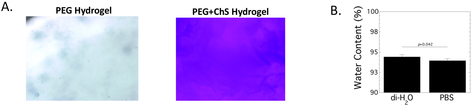
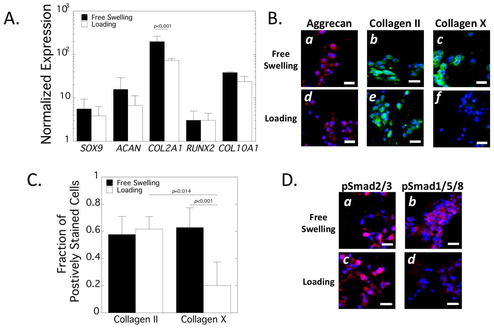
Mesenchymal stem cells (MSCs) are promising for cartilage regeneration, but readily undergo terminal differentiation. The aim of this study was two-fold: a) investigate physiochemical cues from a cartilage-mimetic hydrogel under dynamic compressive loading on MSC chondrogenesis and hypertrophy and b) identify whether Smad signaling and p38 MAPK signaling mediate hypertrophy during MSC chondrogenesis. Human MSCs were encapsulated in photoclickable poly(ethylene glycol) hydrogels containing chondroitin sulfate and RGD, cultured under dynamic compressive loading or free swelling for three weeks, and evaluated by qPCR and immunohistochemistry. Loading inhibited hypertrophy in the cartilage-mimetic hydrogel indicated by a reduction in pSmad 1/5/8, Runx2, and collagen X proteins, while maintaining chondrogenesis by pSmad 2/3 and collagen II proteins. Inhibiting pSmad 1/5/8 under free swelling culture significantly reduced collagen X protein, similar to the loading condition. Chondroitin sulfate was necessary for load-inhibited hypertrophy and correlated with enhanced S100A4 expression, which is downstream of the osmotic responsive transcription factor NFAT5. Inhibiting p38 MAPK under loading reduced S100A4 expression, and upregulated Runx2 and collagen X protein. Findings from this study indicate that chondroitin sulfate with dynamic loading create physiochemical cues that support MSC chondrogenesis and attenuate hypertrophy through Smad 1/5/8 inhibition and p38 MAPK upregulation.


Figures

Figure 1.
Figure 1.
A. Schematic of the formation of the cartilage-mimetic hydrogel using the photoclickable thiol-norbornene reaction between multi-arm norbornene functionalized monomers and thiol containing crosslinkers and extracellular matrix mimetics of chondroitin sulfate and RGD. B. Experimental design of the three studies performed in this work.
Figure 2.
Figure 2.
Characterization of incorporated thiolated-chondroitin sulfate (ChS) into the PEG hydrogel. A. The incorporation of ChS was confirmed by toluidine blue which stains negatively charged molecules blue. Brightfield microscopy images of PEG only and PEG-ChS hydrogels are shown (100x magnification). B. The water content of PEG-ChS hydrogels swollen in diH2O or phosphate buffered saline (PBS) for 24 hours. Data are reported as mean with standard deviation as error bar (n=10).
Figure 3.
Figure 3.
Assessment of chondrogenesis in the cartilage-mimetic hydrogels under loading. A. Normalized gene expression of encapsulated hMSCs at day 21 of free swelling (black bars) and loading (white bars) culture conditions. Relative expression was normalized to pre-encapsulated hMSCs. Data are mean with standard deviation as error bars (n=3). B. Representative confocal microscopy images for aggrecan (red), collagen II (green) and collagen X (green) at day 21 under free swelling (a-c) and loading (d-f) culture conditions. Nuclei are counterstained blue. Scale bar = 20 μm. C. Quantitative analysis of the fraction of positively stained cells for collagen II and collagen X at day 21 in free swelling (black bars) and loading (white bars) culture condition. Data are mean with standard deviation as error bars (n= 4 for IHC). D. Representative confocal microscopy images for pSmad2/3 (red) and pSmad1/5/8 (red) at day 21 under free swelling (a-b) and loading (c-d) culture conditions. Nuclei are counterstained blue. Scale bar = 20 μm.
Figure 4.
Figure 4.
Assessment of SMAD signaling in the cartilage-mimetic hydrogel under loading and with dorsomorphin, an inhibitor of pSmad1/5/8. A. Representative confocal microscopy images at day 14 and day 21 for pSmad2/3 (red) and pSmad1/5/8 (red) under free swelling (a-b,g-h), loading (c-d, i-j), and free swelling with dorsomorphin (e-f, k-l). Nuclei are counterstained blue. Scale bar = 20 μm. C. Quantitative analysis of pSmad2/3 and pSmad1/5/8 intensity per nuclei at day 14 (black bars) and day 21 (white bars) for the different culture conditions. Data are mean with standard deviation as error bars (n=4 hydrogels). Results from two-way ANOVA with pairwise comparisons are provided.
Figure 5.
Figure 5.
Assessment of chondrogenesis in the cartilage-mimetic hydrogel under loading and with dorsomorphin, an inhibitor of pSmad1/5/8. A. Normalized gene expression of encapsulated hMSCs as a function of culture time for free swelling (circle, black line), loading (square, green line), and free swelling with dorsomorphin (diamond, blue line). Relative expression was normalized to pre-encapsulated hMSCs. Data are mean with standard deviation as error bars (n=3). Results from two-way ANOVA are provided. B. Representative confocal microscopy images at day 21 for collagen II (green), Runx2 (green), and collagen X (green) under free swelling (a,d,g), loading (b,e,h), and free swelling with dorsomorphin (c,f,i). Nuclei are counterstained blue. Scale bar = 20 μm. C. Quantitative analysis of the fraction of positively stained cells for collagen II and collagen X at day 21 in free swelling (black bars) and loading (white bars) culture condition. Data are mean with standard deviation as error bars (n=4). Results from two-way ANOVA with pairwise comparisons are provided.
Figure 6.
Figure 6.
Assessment of chondrogenesis in the cartilage-mimetic hydrogel under loading and with SB203580, an inhibitor of p38 signaling. Representative confocal microscopy images at day 21 for A. pSmad2/3 (red), B. collagen II (green), C. pSmad1/5/8 (red), D. Runx2 (green), and E. collagen X (green). Experimental conditions included with (a,c,e,f) and without (b,d,f,h) chondroitin sulfate (ChS) in the cartilage-mimetic hydrogel, free swelling without (a-b) and with (e-f) SB203580, and loading without (c-d) and with (f-h) SB203580. Nuclei are counterstained blue. Scale bar = 20 μm.
Figure 7.
Figure 7.
Assessment of hyperosmotic genes in the cartilage-mimetic hydrogel under loading and with SB203580, an inhibitor of p38 signaling. A. Schematic of hyperosmolarity effects on NFAT5 transcription factor and its downstream control of S100A4. B. Relative gene expression of encapsulated hMSCs at day 7 and day 21 in cartilage-mimetic hydrogel with and without chondroitin sulfate (ChS), SB203580, and loading. Data are mean with standard deviation as error bars (n=3). Three-way ANOVA results for the main effects are provided for NFAT5 at day 21. Pairwise comparisons are provided for S100A4 at day 21.

References

    1. Farrell E, Both SK, Odörfer KI, Koevoet W, Kops N, O’Brien FJ, de Jong RJB, Verhaar JA, Cuijpers V, Jansen J, Erben RG, van Osch GJ, In-vivo generation of bone via endochondral ossification by in-vitro chondrogenic priming of adult human and rat mesenchymal stem cells, BMC Musculoskelet. Disord 12 (2011) 31. doi: 10.1186/1471-2474-12-31. - DOI - PMC - PubMed
    1. Pelttari K, Winter A, Steck E, Goetzke K, Hennig T, Ochs BG, Aigner T, Richter W, Premature induction of hypertrophy during in vitro chondrogenesis of human mesenchymal stem cells correlates with calcification and vascular invasion after ectopic transplantation in SCID mice, Arthritis Rheum. 54 (2006) 3254–3266. doi: 10.1002/art.22136. - DOI - PubMed
    1. Johnstone B, Hering TM, Caplan AI, Goldberg VM, Yoo JU, In vitro chondrogenesis of bone marrow-derived mesenchymal progenitor cells, Exp. Cell Res 238 (1998) 265–272. doi: 10.1006/excr.1997.3858. - DOI - PubMed
    1. Varghese S, Hwang NS, Canver AC, Theprungsirikul P, Lin DW, Elisseeff J, Chondroitin sulfate based niches for chondrogenic differentiation of mesenchymal stem cells, Matrix Biol. 27 (2008) 12–21. doi: 10.1016/j.matbio.2007.07.002. - DOI - PubMed
    1. Bryant SJ, Davis-Arehart KA, Luo N, Shoemaker RK, Arthur JA, Anseth KS, Synthesis and Characterization of Photopolymerized Multifunctional Hydrogels: Water-Soluble Poly(Vinyl Alcohol) and Chondroitin Sulfate Macromers for Chondrocyte Encapsulation, Macromolecules. 37 (2004) 6726–6733. doi: 10.1021/ma0499324. - DOI

Publication types