. 2019 Jan:20:354-366.
doi: 10.1016/j.redox.2018.10.017. Epub 2018 Oct 26.

A maladaptive ER stress response triggers dysfunction in highly active muscles of mice with SELENON loss

Affiliations

A maladaptive ER stress response triggers dysfunction in highly active muscles of mice with SELENON loss

Diego Pozzer et al. Redox Biol. 2019 Jan.

Abstract

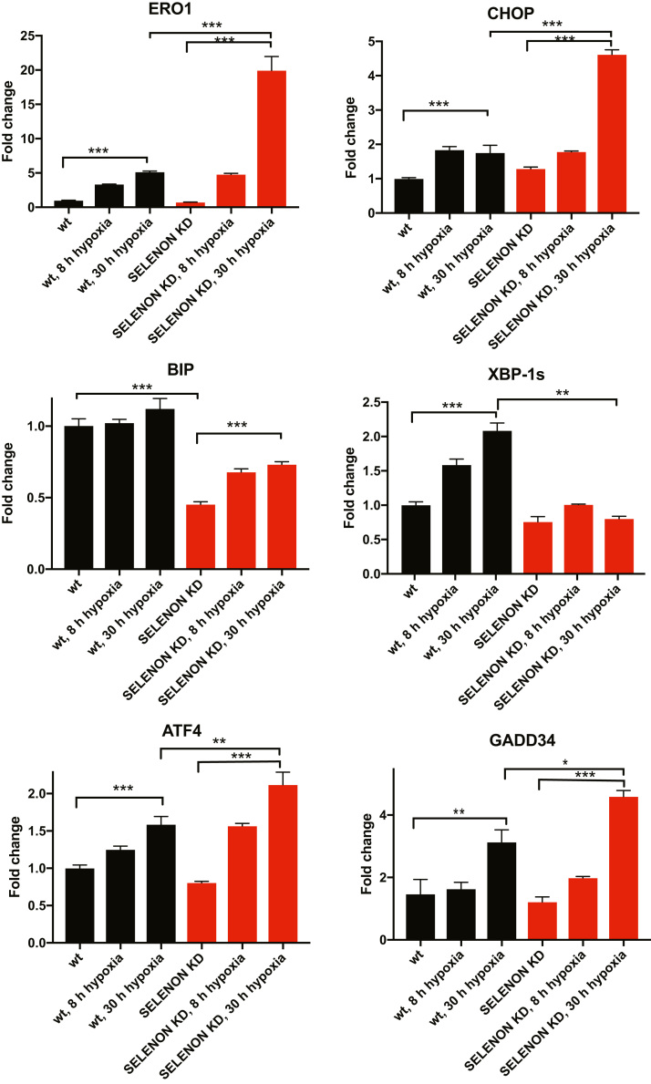
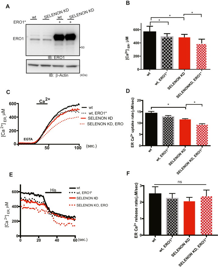
Selenoprotein N (SELENON) is an endoplasmic reticulum (ER) protein whose loss of function leads to human SELENON-related myopathies. SelenoN knockout (KO) mouse limb muscles, however, are protected from the disease, and display no major alterations in muscle histology or contractile properties. Interestingly, we find that the highly active diaphragm muscle shows impaired force production, in line with the human phenotype. In addition, after repeated stimulation with a protocol which induces muscle fatigue, also hind limb muscles show altered relaxation times. Mechanistically, muscle SELENON loss alters activity-dependent calcium handling selectively impinging on the Ca2+ uptake of the sarcoplasmic reticulum and elicits an ER stress response, including the expression of the maladaptive CHOP-induced ERO1. In SELENON-devoid models, ERO1 shifts ER redox to a more oxidised poise, and further affects Ca2+ uptake. Importantly, CHOP ablation in SelenoN KO mice completely prevents diaphragm dysfunction, the prolonged limb muscle relaxation after fatigue, and restores Ca2+ uptake by attenuating the induction of ERO1. These findings suggest that SELENON is part of an ER stress-dependent antioxidant response and that the CHOP/ERO1 branch of the ER stress response is a novel pathogenic mechanism underlying SELENON-related myopathies.

Keywords: Diaphragm dysfunction; ER stress response; SELENON.


Figures

Fig. 1
Fig. 1
Hypoxia elicits an exacerbated ER stress response with a prominent increase in CHOP and its target ERO1 in SELENON depleted myoblasts. A) Semi-quantitative, real-time RT-PCR analysis of ER stress response markers in mRNA prepared from wild-type (WT) and SELENON KD C2C12 cells exposed to hypoxia for the indicated periods of time (hours, h) (n = 4).
Fig. 2
Fig. 2
SELENON defends ER redox poise in presence of ERO1. A) Immunoblot of the ER-localised redox marker protein roGFP1_iE-Flag, FlagM1-immunoprecipitated from extracts of WT and SELENON KD cells, and resolved by means of non-reducing or reducing SDS-PAGE. The positions of the reduced (red) and oxidised (ox) forms of roGFP_iE are indicated in the non-reducing condition. Below, ERO1 immunoblot of the proteins from mock transfected, transfected with roGFP1_iE-Flag and with the bicistronic vector containing ERO1* and roGFP1_iE-Flag. β-Actin was used as a loading control. Quantification (bottom) of the oxidised on reduced form of roGFP1_iE-Flag in non-reducing condition indicates the similar distribution of roGFP1_iE in the reduced and oxidised forms under basal conditions in both cell types, and the higher accumulation of the oxidised form in SELENON KD cells after ERO1* expression (n = 3). B) Immunoblot of endogenous PDI isoforms extracted from WT, SELENON KD, ERO1 KD and double SELENON/ERO1 KD cells after a reductive pulse with dithiothreitol, subsequent three washouts and resolved by means of non-reducing SDS-PAGE. The positions of the reduced and oxidised forms of the protein are indicated; the asterisk marks the position of redox-insensitive proteins that are insensitive to DTT and also react with this antibody (as already seen in . Below, percentage of re-oxidation (with respect to the completely reduced DTT-treated PDI) during the three washouts of PDI. Note the faster re-oxidation of PDI in the double SELENON/ERO1 KD cells indicated by the quantification.
Fig. 3
Fig. 3
Lack of SELENON and high levels of ERO1 coordinatedly affect SERCA activity. A) ERO1 and β-Actin Immunoblots of WT and SELENON KD HeLa cells overexpressing ERO1*(C104A, C131A). B) WT and SELENON KD HeLa cells were co-transfected with ER aequorin and ERO1* and Ca2+ refilling of the ER was recorded. Bar graphs representing steady-state ER Ca2+ concentrations (n = 8). C) Measurements of ER [Ca2+] refilling. The traces are representative of eight independent experiments that gave similar results. D) Bar graphs representing the rate of Ca2+ uptake in the ER (n = 8). E) Measurements of ER [Ca2+] efflux after agonist stimulation (Histamin 100 mM). The traces are representative of eight independent experiments that gave similar results. F) Bar graphs representing measurements of ER [Ca2+] efflux after agonist stimulation (n = 8) (ns stands for not statistically significant).
Fig. 4
Fig. 4
Lack of SELENON increases half-relaxation time by affecting SERCA activity. A) Half-relaxation time (milliseconds) in WT and SELENON KO muscle fibres from FDB muscle upon electrical stimulation with single twitches. B) Example of traces of caffeine-induced Ca2+ release in wildtype and SELENON KO fibres during electrical stimulation with single twitches. C) Maximal caffeine-induced Ca2+ release in WT and SELENON KO muscle fibres from FDB muscle in electrically stimulated fibres. D) Immunoblot for ERO1 and SERCA2 in WT and SELENON KO fibres mock-transduced or transduced with AAV-SERCA2A and AAV-ERO1. On the right, ERO1 and SERCA2 protein quantification after AAV infection. E) Half relaxation time in WT and SELENON KO fibres mock-transduced or transduced with AAV-SERCA2A and AAV-ERO1 upon electrical stimulation.
Fig. 5
Fig. 5
Diaphragm dysfunction in SELENON KO is associated with an exacerbated ER stress response. A) Semi-quantitative, real-time RT-PCR analysis of ER stress response markers in mRNA prepared from WT and SELENON KO diaphragms of 4- and 24-week-old mice (n = 12 for diaphragm of 4-week-old mice, n = 6 for diaphragms of 24-week-old). Bottom: ERO1 and BIP immunoblots and relative quantifications of the signals of proteins from 24-week-old mice, GAPDH was used as a loading control. B) Representative frequency curve, tetanic force and half relaxation time (stimulation frequency of 100 Hz) measured in vivo in the leg muscles (that mainly represents the force of the gastrocnemius muscle) (n = 12) and measured ex-vivo in strips of diaphragm (n = 20). C) Representative histology of H&E of diaphragms and minimal Feret's diameter (μm) of WT and SELENON KO diaphragms (n = 1200 fibres). Bottom: Representative fiber type immunostaining images in diaphragms using specific myosin heavy chain antibodies (Scale bars are 100 µm).
Fig. 6
Fig. 6
Deleting CHOP rescues diaphragm dysfunction in SELENON KO mice by reducing ERO1 levels. A) Semi-quantitative, real-time RT-PCR analysis of ER stress response markers of mRNA prepared from wild-type (WT) and SELENON KO, CHOP KO and double SELENON, CHOP KO (DKO) diaphragms (n = 8). B) Representative immunoblot of newly synthesised, puromycin-labelled proteins using an anti-puromycin antibody, and bar graphs of their signal in arbitrary units. C) Representative frequency curve and tetanic force measured ex-vivo on strips of diaphragm (n = 8).
Fig. 7
Fig. 7
Deleting CHOP rescues defective limb muscle relaxation in SELENON KO mice by reducing ERO1 levels. A) Relative force reduction in limb muscles before and after 90 repeated maximal tetanic stimulations (n = 6). B) Semi-quantitative real-time RT-PCR analysis of ER stress response markers in mRNA prepared from WT, SELENON KO and DKO soleus of 24-week-old sedentary mice (rest) and mice after one bout of treadmill running (run). C) Half-relaxation time in isolated FDB fibres after electrical stimulation.
Fig. 8
Fig. 8
Maladaptive ER stress response in SELENON KO. The ER stress response is an ancient multi-dimensional signaling pathway initiated by ER stress and activated during muscle activity. Usually the ER stress response helps relieve cells from this stress so it serves as an important pro-survival pathway. However, high levels of ER stress persist if a “maladaptive ER stress response” fails to re-establish ER homeostasis and consequently cells are committed to dysfunction. Here we show that SELENON is part of an ER stress-dependent antioxidant response which, if it is missing, makes the CHOP/ERO1 branch of the ER stress response maladaptive quite likely by oxidizing and inhibiting SERCA2 in the highly active muscles.

References

    1. Acosta-Alvear D., Zhou Y., Blais A., Tsikitis M., Lents N.H., Arias C., Lennon C.J., Kluger Y., Dynlacht B.D. XBP1 controls diverse cell type- and condition-specific transcriptional regulatory networks. Mol. Cell. 2007;27:53–66. - PubMed
    1. Allen D.G., Westerblad H. The effects of caffeine on intracellular calcium, force and the rate of relaxation of mouse skeletal muscle. J. Physiol. 1995;487(Pt 2):331–342. - PMC - PubMed
    1. Arbogast S., Ferreiro A. Selenoproteins and protection against oxidative stress: selenoprotein N as a novel player at the crossroads of redox signaling and calcium homeostasis. Antioxid. Redox Signal. 2010;12:893–904. - PubMed
    1. Blaauw B., Canato M., Agatea L., Toniolo L., Mammucari C., Masiero E., Abraham R., Sandri M., Schiaffino S., Reggiani C. Inducible activation of Akt increases skeletal muscle mass and force without satellite cell activation. FASEB J. 2009;23:3896–3905. - PubMed
    1. Bonora M., Giorgi C., Bononi A., Marchi S., Patergnani S., Rimessi A., Rizzuto R., Pinton P. Subcellular calcium measurements in mammalian cells using jellyfish photoprotein aequorin-based probes. Nat. Protoc. 2013;8:2105–2118. - PubMed

Publication types

MeSH terms