Review
. 2018 Dec;10(6):1553-1569.
doi: 10.1007/s12551-018-0468-6. Epub 2018 Nov 3.

Mechanisms of formin-mediated actin assembly and dynamics

Affiliations
Review

Mechanisms of formin-mediated actin assembly and dynamics

Naomi Courtemanche. Biophys Rev. 2018 Dec.

Abstract

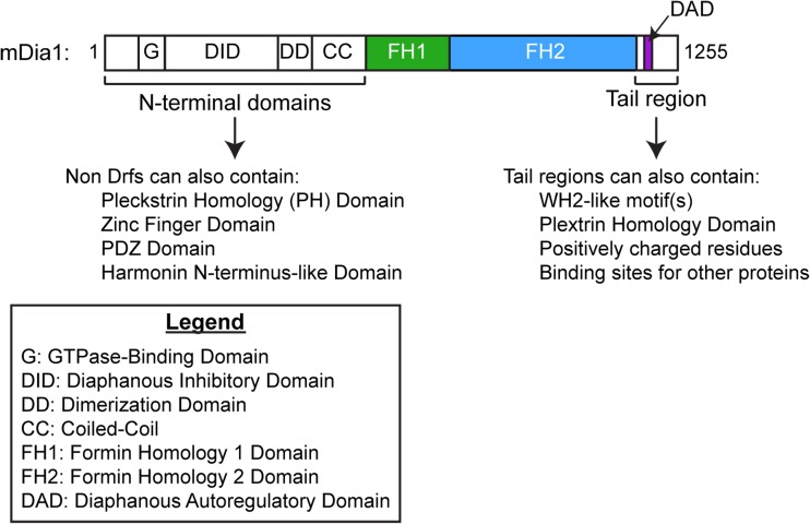
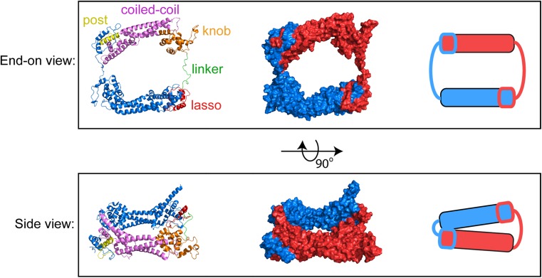
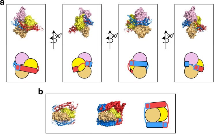
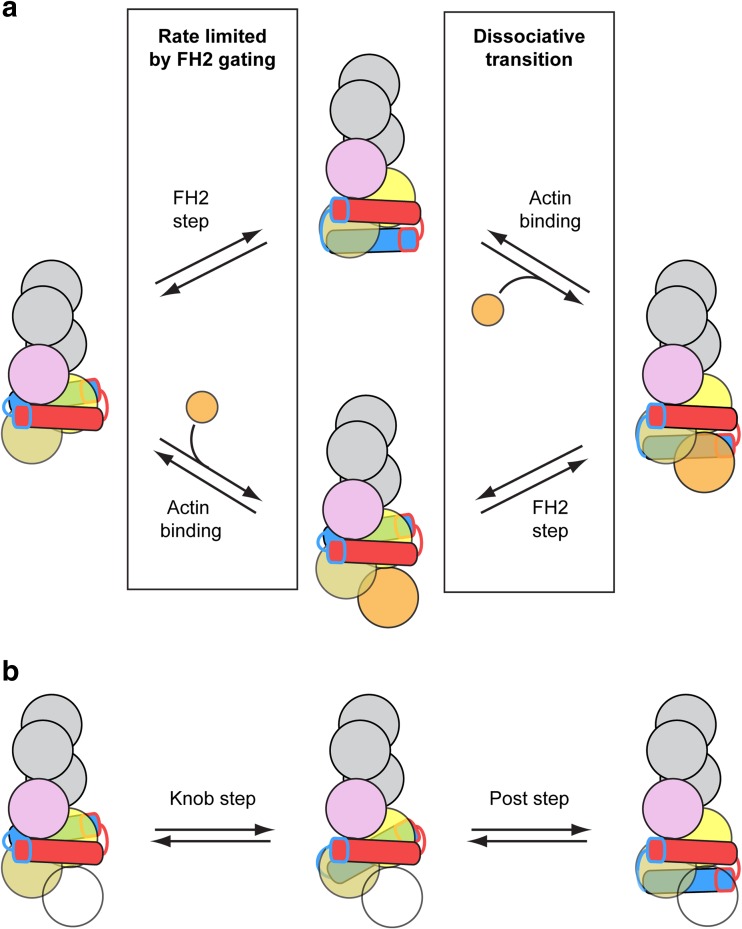
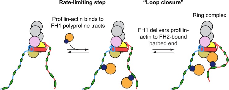
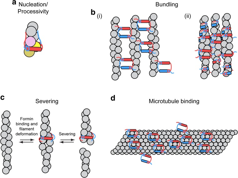
Cellular viability requires tight regulation of actin cytoskeletal dynamics. Distinct families of nucleation-promoting factors enable the rapid assembly of filament nuclei that elongate and are incorporated into diverse and specialized actin-based structures. In addition to promoting filament nucleation, the formin family of proteins directs the elongation of unbranched actin filaments. Processive association of formins with growing filament ends is achieved through continuous barbed end binding of the highly conserved, dimeric formin homology (FH) 2 domain. In cooperation with the FH1 domain and C-terminal tail region, FH2 dimers mediate actin subunit addition at speeds that can dramatically exceed the rate of spontaneous assembly. Here, I review recent biophysical, structural, and computational studies that have provided insight into the mechanisms of formin-mediated actin assembly and dynamics.

Keywords: Actin; Formin; Polymerization; Profilin.


Conflict of interest statement

Conflict of interest

Naomi Courtemanche declares that she has no conflict of interest.

Ethical approval

This article does not contain any studies with human participants or animals performed by the authors.

Figures

Fig. 1
Fig. 1
Domain architecture of mDia1, a diaphanous-related formin (Drf). Domain boundaries are shown to scale. The three domains that influence actin dynamics are the FH1 domain, FH2 domain, and tail region
Fig. 2
Fig. 2
Atomic structure of the dimeric FH2 domain of Bni1p. End-on and side views of the dimeric FH2 domain of the S. cerevisiae formin Bni1p. The structure depicts the actin-bound FH2 dimer, based on pdb ID 1Y64 (Otomo et al. 2005), following 160 ns of all-atom molecular dynamics simulations (Baker et al. 2015). Left = Ribbon representations of the FH2 dimer. One monomer is color-coded to highlight the lasso, linker, knob, coiled-coil and post subdomains. The second monomer is depicted in blue. Center = surface representations of the FH2 dimer; the monomers are depicted in red and blue. Right = cartoon representations of the FH2 dimer
Fig. 3
Fig. 3
Structure of the dimeric FH2 domain of Bni1p bound to actin. Structural representations of the FH2 dimer of the S. cerevisiae formin Bni1p bound to the three terminal subunits of an actin filament following 160 ns of all-atom molecular dynamics simulations (Baker et al. 2015). The initial structural model was constructed based on the crystal structure of the actin-bound Bni1p FH2 domain (pdb ID 1Y64) (Otomo et al. 2005). The structures of the FH2 domains are represented as ribbon diagrams and the individual domains are colored red and blue. The structures of the three actin subunits are depicted as surface representations. The terminal, barbed end subunit is depicted in light orange. The other two actins are shown in yellow and pink. a Multiple orientations of the side view are depicted. The FH2 dimer forms contacts with all three actin subunits. Cartoon representations are also shown to orient the reader. b End-on view of the FH2-actin complex. The FH2 dimer is shown both with ribbon diagrams (right) and as a surface representation (center). A cartoon representation is also shown (left)
Fig. 4
Fig. 4
Two models for FH2-mediated actin polymerization. a Cartoon representation of the addition of an actin subunit (orange circle) to the barbed end of an actin filament bound by an FH2 dimer. Upper reaction scheme = the “stair-stepping” model in which the trailing FH2 domain (blue domain) steps forward before an incoming actin subunit (orange circle) binds to the barbed end. Lower reaction scheme = the “stepping second” model, in which actin binds to the barbed end prior to stepping of the trailing FH2 domain. In both models, the first reaction is limited by FH2 gating. Dissociation of the FH2 dimer from the barbed end is most likely to occur during the second reaction, which involves the population of a dissociative transition state (Cao et al. ; Paul and Pollard 2008, 2009b). b Cartoon representation of a two-step model for the translocation of the trailing FH2 domain, in which an intermediate state is populated (Thompson et al. 2013). The intermediate state is formed by dissociation of the knob subdomain from its trailing binding site (pink circle), followed by translocation and association with a binding site at the barbed end (light orange circle). In this state, the FH2 dimer is bound to two actin subunits. Complete stepping of the trailing FH2 domain is achieved via subsequent translocation of the post subdomain, as depicted in the second reaction. This two-step model for stepping can occur as part of either the stair-stepping or stepping second model for actin subunit addition. To account for this, the incoming actin subunit is depicted as a transparent circle
Fig. 5
Fig. 5
Model for FH1-mediated delivery of profilin actin to the FH2-bound barbed end. Cartoon representation of the addition of a profilin-bound actin subunit (dark blue and orange circles) to the barbed end of an actin filament bound by a representative formin FH1FH2 construct. The unstructured FH1 domains extend outwards from the FH2 dimer and contain multiple polyproline tracts (in this case, six green ovals per FH1 domain). The number of prolines in each polyproline tract tends to increase with the distance from the FH2 domain. Profilin-actin complexes bind the polyproline tracts and are delivered directly to the barbed end in a “loop closure” reaction via diffusion of the FH1 domain and formation of a ring complex (Vavylonis et al. 2006). Subsequent profilin and polyproline tract dissociation events occur rapidly and are not depicted
Fig. 6
Fig. 6
Formin tail-mediated interactions with actin and microtubules. Cartoon representations of interactions between formin FH2-tail constructs with actin filaments and microtubules. Tail regions are depicted as unstructured regions and are color-coded to match their corresponding FH2 domain. a Electrostatic interactions between tail regions and actin promote formin-mediated filament nucleation and FH2 processivity. b Formins can associate with the sides of actin filaments (i) via binding sites on the exterior surfaces of their FH2 domains or (ii) by encircling the filaments with their FH2 domains. In several cases, actin filament decoration by formins promotes filament bundling. Binding is often strengthened via interactions between formin tail regions and actin. c A model for INF2-mediated severing in which an FH2-tail construct of the formin INF2 encircles an actin filament and creates a localized deformation, which promotes severing (Gurel et al. 2014). d FH2-tail constructs of a representative formin decorate a microtubule (gray tube) via interactions mediated by the post subdomains and strengthened by the tail regions

References

    1. Ahuja R, et al. Cordon-bleu is an actin nucleation factor and controls neuronal morphology. Cell. 2007;131(2):337–350. doi: 10.1016/j.cell.2007.08.030. - DOI - PMC - PubMed
    1. Arimura T, et al. Dilated cardiomyopathy-associated FHOD3 variant impairs the ability to induce activation of transcription factor serum response factor. Circ J. 2013;77(12):2990–2996. doi: 10.1253/circj.CJ-13-0255. - DOI - PubMed
    1. Aydin F, Courtemanche N, Pollard TD, Voth GA. Gating mechanisms during actin filament elongation by formins. Elife. 2018;7:e37342. doi: 10.7554/eLife.37342. - DOI - PMC - PubMed
    1. Baker JL, Courtemanche N, Parton DL, McCullagh M, Pollard TD, Voth GA. Electrostatic interactions between the Bni1p formin FH2 domain and actin influence actin filament nucleation. Structure. 2015;23(1):68–79. doi: 10.1016/j.str.2014.10.014. - DOI - PMC - PubMed
    1. Bartolini F, Gundersen GG. Formins and microtubules. Bba-Mol Cell Res. 2010;1803(2):164–173. doi: 10.1016/j.bbamcr.2009.07.006. - DOI - PMC - PubMed