

Real-time three-dimensional echocardiographic left heart parameters in healthy indian adults
- PMID: 30392502
- PMCID: PMC6204452
- DOI: 10.1016/j.ihj.2017.11.018
Real-time three-dimensional echocardiographic left heart parameters in healthy indian adults
Abstract
Objective: Cardiac chamber dimensions are race and anthropometry dependent. We determined the age and gender specific 3-Dimensional echocardiographic (3DE) reference values for dimensions and function of left ventricle (LV) and left atrium (LA) in normal Indian adults.
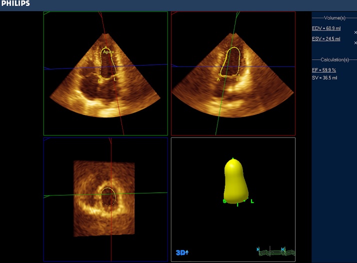
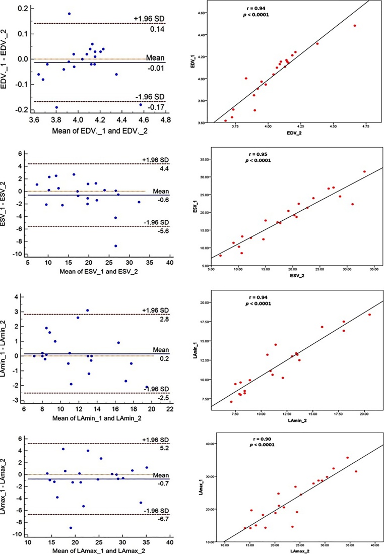
Methods: This single center prospective study enrolled 133 adult Indians free of heart disease and/or hypertensions, subjecting them to 3DE measurements of left atrial (LA) & left ventricular (LV) volumes, function and left ventricular mass (LVM). The higher limits of normal cut-offs were determined for these parameters and their dependency on age, gender and anthropometry were analyzed.
Results: The body surface area (BSA) corrected higher limit cut-offs were: 59.37ml/m2 for LV end diastolic volume (59.19ml/m2 and 59.61ml/m2 for men and women, respectively; P=NS); 23.48ml/m2 for LV end systolic volume (23.27ml/m2 and 23.11ml/m2 for men and women, P=NS). Mean LVEF was 64.79%±7.26 (62.99%±6.51 and 67.05%±7.58 in men and women, P=NS). Men had higher LVM than women (119.79g±23.95 vs. 103.26g±23.76, P<0.001), this difference disappeared after BSA indexing. The higher limit cut-offs for normal LA volumes were 20.49ml for minimum volume (21.18ml and 19.46ml for men and women, P=NS) and 39.76ml for maximum volume (39.60ml and 40.03ml in men and women, P=NS). The parameters were smaller compared to western populations but the differences attenuated after BSA indexing.
Conclusions: The study reports normal 3DE parameters of size and function of left heart chambers in Indians.
Keywords: Left atrial emptying fraction; Left atrial volume; Left ventricular end diastolic volume; Left ventricular end systolic volume; Three-dimensional echocardiography.
Copyright © 2017 Cardiological Society of India. Published by Elsevier B.V. All rights reserved.
Figures
References
-
- Juillière Y., Barbier G., Feldmann L. Additional predictive value of both left and right ventricular ejection fractions on long-term survival in idiopathic dilated cardiomyopathy. Eur Heart J. 1997;18:276–280. - PubMed
-
- Knauth A.L., Gauvreau K., Powell A.J. Ventricular size and function assessed by cardiac MRI predict major adverse clinical outcomes late after tetralogy of Fallot repair. Heart. 2008;94:211–216. - PubMed
-
- Fukuda S., Watanabe H., Daimon M. Normal values of real-time 3-dimensional echocardiographic parameters in a healthy Japanese population: the JAMP-3D Study. Circ J. 2012;76(5):1177–1181. - PubMed
-
- Maffessanti F., Muraru D., Esposito R. Age-, body size-, and sex-specific reference values for right ventricular volumes and ejection fraction by three-dimensional echocardiography: a multicenter echocardiographic study in 507 healthy volunteers. Circ Cardiovasc Imaging. 2013;6(September (5)):700–710. - PubMed
-
- Hinderliter A.L., Light K.C., Willis P.W., 4th Racial differences in left ventricular structure in healthy young adults. Am J Cardiol. 1992;69:1196–1199. - PubMed
MeSH terms
LinkOut - more resources
Full Text Sources
Miscellaneous
