. 2018 Dec 19;100(6):1504-1512.e4.
doi: 10.1016/j.neuron.2018.10.020. Epub 2018 Nov 1.

Scale-Invariant Visual Capabilities Explained by Topographic Representations of Luminance and Texture in Primate V1

Affiliations

Scale-Invariant Visual Capabilities Explained by Topographic Representations of Luminance and Texture in Primate V1

Giacomo Benvenuti et al. Neuron. .

Erratum in

Abstract

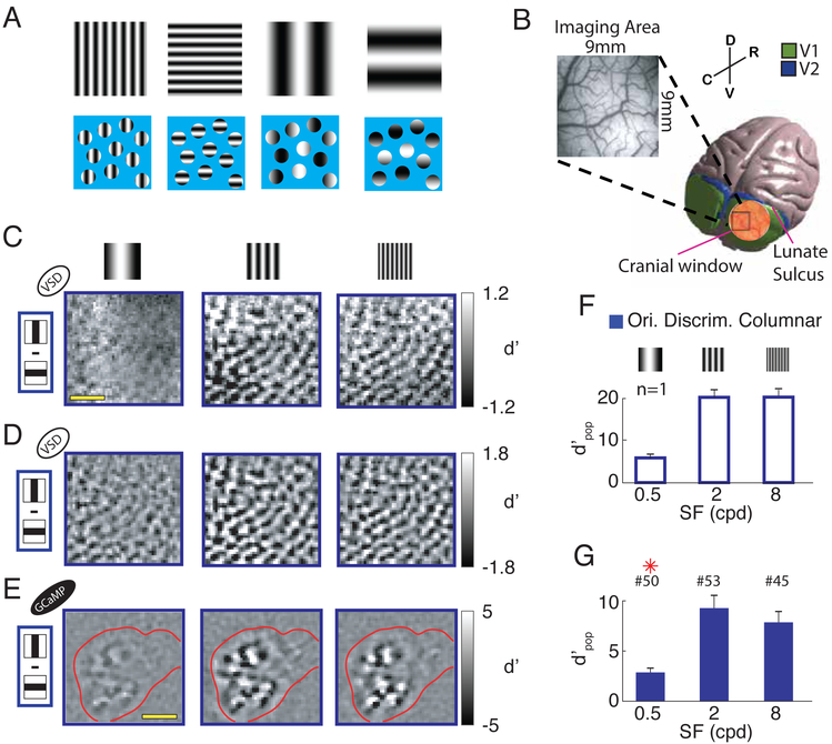
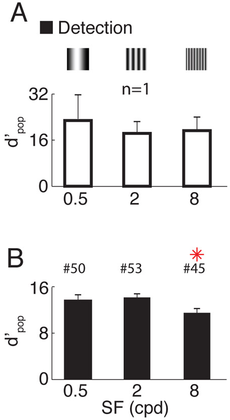
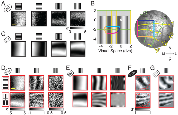
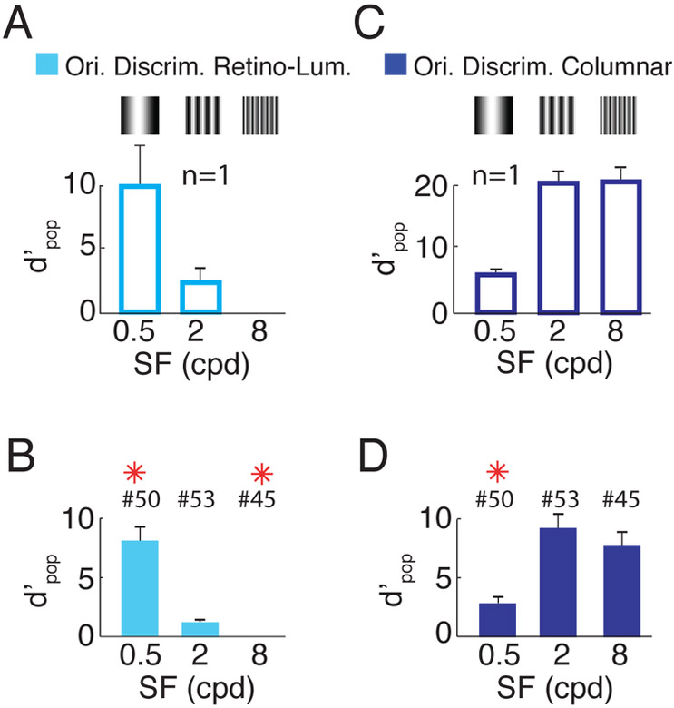
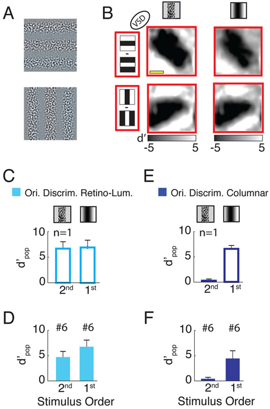
Humans have remarkable scale-invariant visual capabilities. For example, our orientation discrimination sensitivity is largely constant over more than two orders of magnitude of variations in stimulus spatial frequency (SF). Orientation-selective V1 neurons are likely to contribute to orientation discrimination. However, because at any V1 location neurons have a limited range of receptive field (RF) sizes, we predict that at low SFs V1 neurons will carry little orientation information. If this were the case, what could account for the high behavioral sensitivity at low SFs? Using optical imaging in behaving macaques, we show that, as predicted, V1 orientation-tuned responses drop rapidly with decreasing SF. However, we reveal a surprising coarse-scale signal that corresponds to the projection of the luminance layout of low-SF stimuli to V1's retinotopic map. This homeomorphic and distributed representation, which carries high-quality orientation information, is likely to contribute to our striking scale-invariant visual capabilities.

Keywords: genetically encoded cacium indicators; multi-scale representation; neural population code; optical imaging; orientation discrimination; orientation map; primary visual cortex; retinotopic map; visual perception; voltage sensitive dye.


Figures

Figure 1.
Figure 1.. Discriminability of columnar-scale orientation tuned responses to orthogonal gratings as a function of spatial-frequency.
(A) Schematic demonstration of the “aperture problem” for low SF stimuli. The four gratings on the top row (two orientations for medium and low SFs, respectively) are re-displayed in the bottom row behind a blue mask, with small circular apertures, each one representing the RF of a V1 neuron. At low SFs, the oriented contrast pattern in each individual aperture carries limited information about orientation. However, orientation information can be obtained by combining information about the relative positions of apertures with bright and dark patterns. (B) 3D model of the macaque brain with a superimposed image of the cranial window (in scale). The insert shows a picture of the vasculature in an imaging area. Spatial reference abbreviations: R=rostral, C=caudal, D=dorsal, V=ventral. (C) Pixel-by-pixel discriminability (d’ map) across responses to two orthogonal gratings, for three stimulus SFs (0.5, 2 and 8cpd), from an example session. Amplitude expressed in signed d′. (D) Similar to C but isolating the columnar response component with bandpass filtration at 0.8-3 cycles per mm. (E)Similar to D but in a different monkey, using calcium imaging. Red contour represents cortical area in which the calcium indicator GCaMP6f is expressed. Scale bar in C and E - 2mm. (F) Overall discriminability (d′pop; see Methods) of columnar responses to gratings stimuli with orthogonal orientations, at the three SFs, in the same example session as in C and D. Error-bars indicate bootstrapped standard errors. (G) Similar to F but summary across sessions. Error-bars indicate standard-error of the mean across sessions. Red asterisks signal significant difference with respect to the central SF condition (two-sample t-test P<0.05). The number of sessions per SF is indicated above the corresponding bar. All figures and text refer to VSD imaging unless explicitly noted otherwise. Ori. Discrim – orientation discrimination. Here and in subsequent figures, open bars indicate results from an example session while solid bars indicate summary across sessions.
Figure 2.
Figure 2.. Detectability of responses to grating stimuli as a function of stimulus spatial frequency.
(A) Overall discriminability between responses to gratings vs. blank, at the three different stimulus SFs, in the same example session as in Figure 1. Error-bars indicate bootstrapped standard errors. (B) Similar to A but summary across sessions. Error-bars indicate standard-error of the mean across sessions. Red asterisks signal significant difference with respect to the central SF condition (two-sample t-test, P<0.05). The number of sessions per SF is indicated above the corresponding bar.
Figure 3.
Figure 3.. Layout of luminance-retinotopic responses to grating stimuli as a function of stimulus orientation, phase and spatial-frequency.
(A) Each panel displays the difference (in signed d′) between neural responses to a low SF (0.5 cpd) grating stimulus and a blank stimulus. The stimuli are the four combinations of two opposite phases and two orthogonal orientations conditions (see cartoon on top). Same example session as in Figure 1. (B) Representation of visual space in the imaged area. Left - The central white dot indicates the fixation point. Bottom-left – 0.5 cpd grating. The colored outline indicates the projection to visual space of the colored rectangle on the right panel (which represents the imaged area). The yellow and green lines represent visual space coordinates. Right - Image of the cortex within the cranial window with retinotopic coordinates superimposed. Brain orientation spatial references: A=anterior, P=posterior, M=medial, L=lateral. The V1/V2 border is located several mm anterior to the imaged area and is approximately parallel to the dark blue edge of the colored square. Consistently with previous studies (e.g., Blasdel and Campbell, 2001), cortical magnification in the direction parallel to the V1/V2 border (and normal to ocular dominance columns in this regions of V1) is much larger than in the direction normal to the V1/V2 border (and parallel to ocular dominance columns). This is why the projection of the square from the cortex to the visual field is strongly anisotropic. (C) Simulation (Sim) of the luminance-retinotopic signal presented in A under the assumption of stronger V1 population response to the dark stimulus regions. The average correlation between the predicted and observed patterns is 0.80. Because the prediction under the assumption of bright dominance is of opposite polarity to the prediction under the assumption of dark dominance, the average correlation between the observed response and the one predicted under a bright-dominance assumption is equal to, and of opposite sign, to the correlation with the prediction under a dark-dominance assumption. (D) Mean differences in neural responses divided by their standard deviations (signed d’ values) to grating stimuli with opposite phase angles, for the two orthogonal orientations (rows) and the three SFs (columns). (E) Simulation of the luminance-retinotopic signal presented in D. Average correlation between the predicted and observed pattern is 0.93 at 0.5 cpd, 0.39 at 2 cpd and 0.02 at 8 cpd. (F) Similar to D but in another example experiment and only for the medium SF condition (2cpd), using calcium imaging. Red contour represents cortical area in which the calcium indicator is expressed. (G) Simulation of the luminance-retinotopic signal presented in F. Average correlation between the predicted and observed patterns is 0.62. Scale bar in A, D and F - 2mm. Note that because of the anisotropy in the retinotopic map, the spatial frequency of the predicted pattern is higher for vertical than for horizontal stimuli.
Figure 4.
Figure 4.. Discriminability based on luminance-retinotopic and columnar responses to orthogonal gratings as a function of spatial-frequency.
(A) Overall discriminability based on luminance-retinotopic signals between grating stimuli with orthogonal orientations, at the three SFs, for the same example session as in Figures 1 and 2. Error-bars indicate bootstrapped standard errors. (B) Similar to A but averaged across sessions. Same format as Figure 1G. The bars and error bars for 8 cpd in A and B are too small to see. (C,D). Same as A,B but for columnar signals (reproduced from Figure 1F,G). See Figure S1 and S4 for results from individual monkeys. Ori. Discrim – orientation discrimination. Retino-Lum – luminance-retinotopic.
Figure 5.
Figure 5.. Discriminability of neural responses to second- and first-order gratings with orthogonal orientations.
(A) Example of second order gratings stimuli with orthogonal orientations (the amplitude is clipped for display purposes). (B) Difference (d′ map) across responses to two low SF (0.5 cpd) gratings stimuli with opposite phases for the two orthogonal orientations (rows). The two columns represent responses to second and first order stimuli respectively when the region with the high contrast in the second order stimulus overlaps the dark region in the first order stimulus. The average correlation between the responses to first and second order stimuli is 0.96. (C) Discriminability of the luminance-retinotopic (cyan) responses to second and first order stimuli with orthogonal orientations and low SF (same session as in B). (D) Similar to C but averaged across 6 sessions. (E,F) Similar to C and D but for the columnar signals. Scale bar in B - 2mm.

Similar articles

Cited by

References

    1. Adelson E, and Movshon J (1982). Phenomenal coherence of moving visual patterns. Nature 300, 523–525. - PubMed
    1. Adelson EH, and Bergen JR (1985). Spatiotemporal energy models for the perception of motion. J. Opt. Soc. Am. A 2, 284–299. - PubMed
    1. Angelucci A, Levitt JB, and Lund JS (2002). Anatomical origins of the classical receptive field of single neurons in macaque visual cortical area V1. Prog. Brain Res 136, 373–388. - PubMed
    1. Blasdel GG (1992). Orientation Selectivity, Striate Cortex Preference, and Continuity in Monkey. J. Neurosci 12, 3139–3161. - PMC - PubMed
    1. Blasdel GG, and Campbell D (2001). Functional retinotopy of monkey visual cortex. J. Neurosci 21, 8286–8301. - PMC - PubMed

Publication types