

Persistence of Systemic Murine Norovirus Is Maintained by Inflammatory Recruitment of Susceptible Myeloid Cells
- PMID: 30392829
- PMCID: PMC6248887
- DOI: 10.1016/j.chom.2018.10.003
Persistence of Systemic Murine Norovirus Is Maintained by Inflammatory Recruitment of Susceptible Myeloid Cells
Abstract
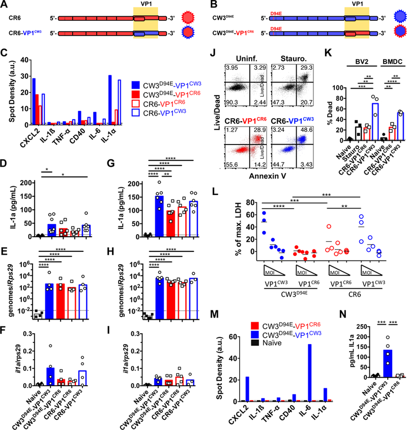
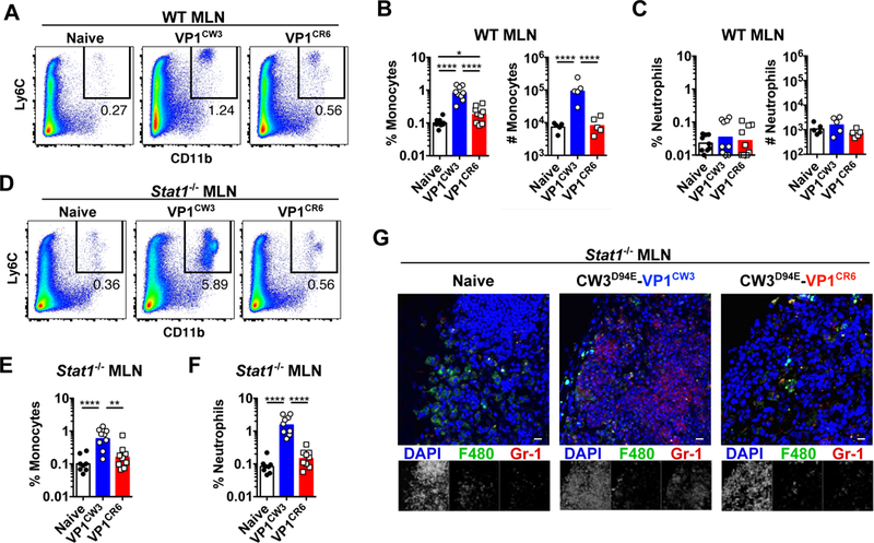
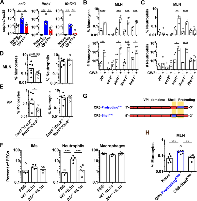
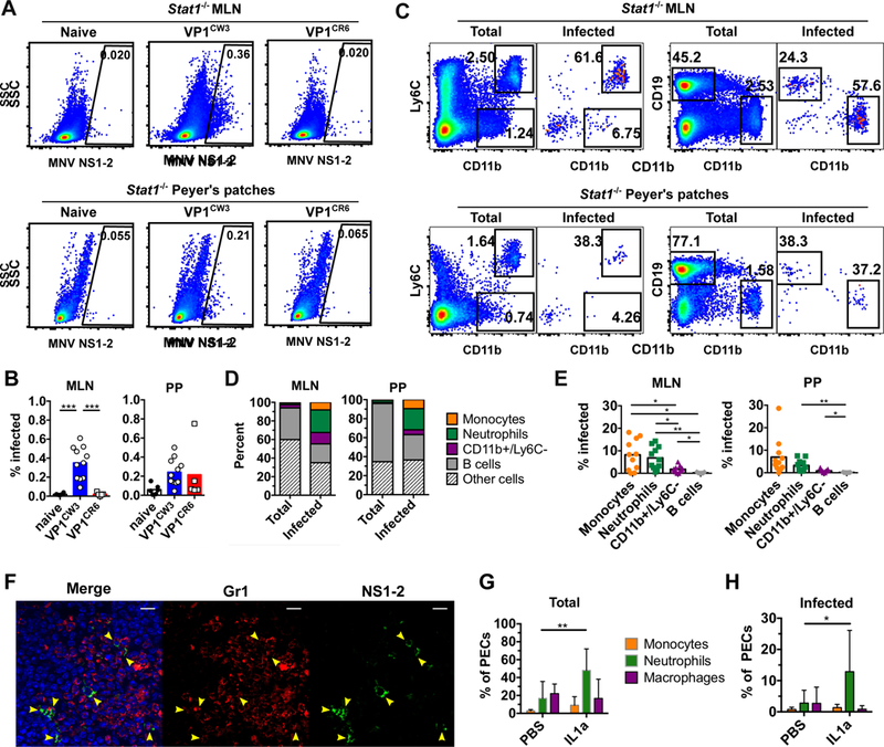
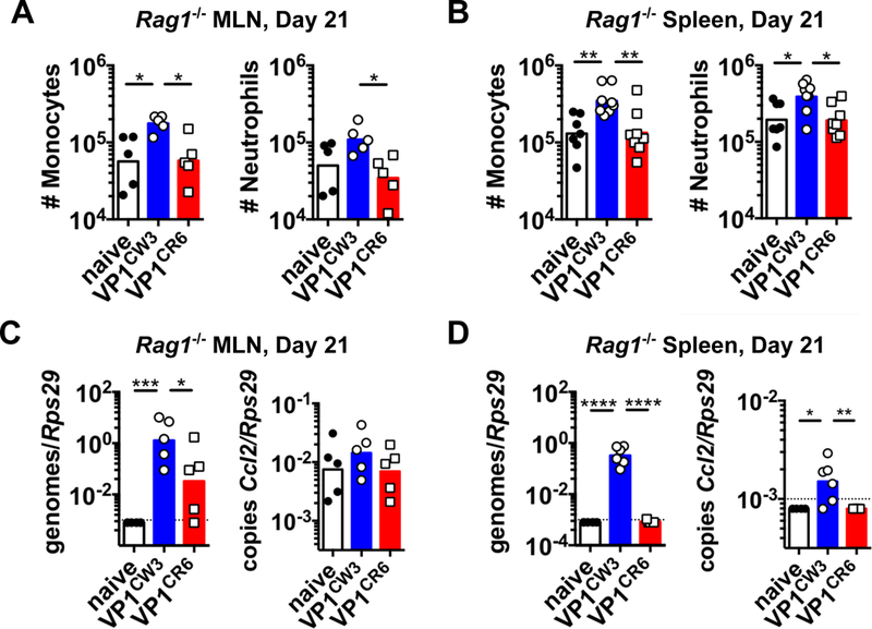
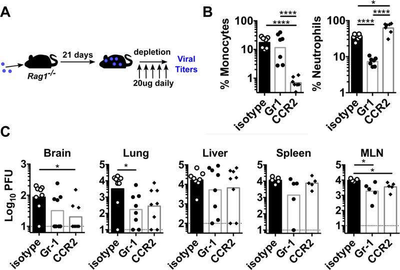
Viral persistence can contribute to chronic disease and promote virus dissemination. Prior work demonstrated that timely clearance of systemic murine norovirus (MNV) infection depends on cell-intrinsic type I interferon responses and adaptive immunity. We now find that the capsid of the systemically replicating MNV strain CW3 promotes lytic cell death, release of interleukin-1α, and increased inflammatory cytokine release. Correspondingly, inflammatory monocytes and neutrophils are recruited to sites of infection in a CW3-capsid-dependent manner. Recruited monocytes and neutrophils are subsequently infected, representing a majority of infected cells in vivo. Systemic depletion of inflammatory monocytes or neutrophils from persistently infected Rag1-/- mice reduces viral titers in a tissue-specific manner. These data indicate that the CW3 capsid facilitates lytic cell death, inflammation, and recruitment of susceptible cells to promote persistence. Infection of continuously recruited inflammatory cells may be a mechanism of persistence broadly utilized by lytic viruses incapable of establishing latency.
Keywords: inflammation; monocytes; neutrophils; norovirus; persistence.
Copyright © 2018 Elsevier Inc. All rights reserved.
Conflict of interest statement
Declaration of Interests
We declare no competing interests.
Figures
Comment in
-
A pLOT of Viral Persistence.Cell Host Microbe. 2018 Nov 14;24(5):618-619. doi: 10.1016/j.chom.2018.10.010. Cell Host Microbe. 2018. PMID: 30439337
Similar articles
-
Persistent enteric murine norovirus infection is associated with functionally suboptimal virus-specific CD8 T cell responses.J Virol. 2013 Jun;87(12):7015-31. doi: 10.1128/JVI.03389-12. Epub 2013 Apr 17. J Virol. 2013. PMID: 23596300 Free PMC article.
-
Type I Interferon Receptor Deficiency in Dendritic Cells Facilitates Systemic Murine Norovirus Persistence Despite Enhanced Adaptive Immunity.PLoS Pathog. 2016 Jun 21;12(6):e1005684. doi: 10.1371/journal.ppat.1005684. eCollection 2016 Jun. PLoS Pathog. 2016. PMID: 27327515 Free PMC article.
-
Comparative murine norovirus studies reveal a lack of correlation between intestinal virus titers and enteric pathology.Virology. 2011 Dec 20;421(2):202-10. doi: 10.1016/j.virol.2011.09.030. Epub 2011 Oct 22. Virology. 2011. PMID: 22018636 Free PMC article.
-
Norovirus encounters in the gut: multifaceted interactions and disease outcomes.Mucosal Immunol. 2019 Nov;12(6):1259-1267. doi: 10.1038/s41385-019-0199-4. Epub 2019 Sep 9. Mucosal Immunol. 2019. PMID: 31501514 Free PMC article. Review.
-
The Role of Interferon in Persistent Viral Infection: Insights from Murine Norovirus.Trends Microbiol. 2018 Jun;26(6):510-524. doi: 10.1016/j.tim.2017.10.010. Epub 2017 Nov 17. Trends Microbiol. 2018. PMID: 29157967 Free PMC article. Review.
Cited by
-
Interferons and tuft cell numbers are bottlenecks for persistent murine norovirus infection.PLoS Pathog. 2024 May 3;20(5):e1011961. doi: 10.1371/journal.ppat.1011961. eCollection 2024 May. PLoS Pathog. 2024. PMID: 38701091 Free PMC article.
-
Interferon regulatory factor 6 (IRF6) determines intestinal epithelial cell development and immunity.Mucosal Immunol. 2024 Aug;17(4):633-650. doi: 10.1016/j.mucimm.2024.03.013. Epub 2024 Apr 9. Mucosal Immunol. 2024. PMID: 38604478 Free PMC article.
-
Characterizing a Murine Model for Astrovirus Using Viral Isolates from Persistently Infected Immunocompromised Mice.J Virol. 2019 Jun 14;93(13):e00223-19. doi: 10.1128/JVI.00223-19. Print 2019 Jul 1. J Virol. 2019. PMID: 30971471 Free PMC article.
-
Nlrp3 inflammasome activation and Gasdermin D-driven pyroptosis are immunopathogenic upon gastrointestinal norovirus infection.PLoS Pathog. 2019 Apr 24;15(4):e1007709. doi: 10.1371/journal.ppat.1007709. eCollection 2019 Apr. PLoS Pathog. 2019. PMID: 31017981 Free PMC article.
-
Tuft-cell-intrinsic and -extrinsic mediators of norovirus tropism regulate viral immunity.Cell Rep. 2022 Nov 8;41(6):111593. doi: 10.1016/j.celrep.2022.111593. Cell Rep. 2022. PMID: 36351394 Free PMC article.
References
Publication types
MeSH terms
Substances
Grants and funding
LinkOut - more resources
Full Text Sources
Medical
Molecular Biology Databases
