. 2018 Nov 15;175(5):1228-1243.e20.
doi: 10.1016/j.cell.2018.10.006. Epub 2018 Nov 1.

N6-methyladenine DNA Modification in Glioblastoma

Affiliations

N6-methyladenine DNA Modification in Glioblastoma

Qi Xie et al. Cell. .

Abstract

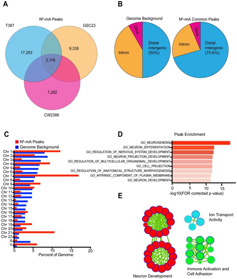
Genetic drivers of cancer can be dysregulated through epigenetic modifications of DNA. Although the critical role of DNA 5-methylcytosine (5mC) in the regulation of transcription is recognized, the functions of other non-canonical DNA modifications remain obscure. Here, we report the identification of novel N6-methyladenine (N6-mA) DNA modifications in human tissues and implicate this epigenetic mark in human disease, specifically the highly malignant brain cancer glioblastoma. Glioblastoma markedly upregulated N6-mA levels, which co-localized with heterochromatic histone modifications, predominantly H3K9me3. N6-mA levels were dynamically regulated by the DNA demethylase ALKBH1, depletion of which led to transcriptional silencing of oncogenic pathways through decreasing chromatin accessibility. Targeting the N6-mA regulator ALKBH1 in patient-derived human glioblastoma models inhibited tumor cell proliferation and extended the survival of tumor-bearing mice, supporting this novel DNA modification as a potential therapeutic target for glioblastoma. Collectively, our results uncover a novel epigenetic node in cancer through the DNA modification N6-mA.

Keywords: DNA methylation; H3K9me3; N(6)-methyladenine; brain tumor; cancer stem cell; chromatin; epigenetics; glioblastoma; heterochromatin; neuro-oncology.


Figures

Figure 1.
Figure 1.. Identification of N(6)-methyladenine (N6-mA) DNA modification in human glioblastoma. See also Figure S1.
(A) Levels of the N6-mA DNA modification were assessed via DNA dot blot in (1) normal human astrocytes, (2) patient-derived GSC models (387, D456, GSC23, and 1919) and (3) primary human glioblastoma specimens (3028, CW2386) using an N6-mA-specific antibody. Methylene blue detected DNA loading. (B) Mass spectrometry analysis of N6-mA in two normal human astrocyte cell lines and two patient-derived GSC models (387 and D456). Data are presented as mean ± SD. Two replicates were used for each sample. Significance was determined by one-way ANOVA with Tukey multiple comparison test. P < 0.0001 for each human astrocyte vs. GSC comparison. (C) N6-mA DNA immunofluorescence in normal human astrocytes and human patient-derived GSC models (387, D456, GSC23). DAPI indicates cell nuclei. Scale bars, 50 μm. (D) Quantification of percentage of N6-mA positive cells by immunofluorescence staining in (C). N6-mA was quantified counting 100 cells from each sample. N = 3 slides/cell type. Data are presented as mean ± SD. Significance assessed by ANOVA. ***, P < 0.001. (E) Immunohistochemistry (IHC) staining of N6-mA in non-neoplastic brain tissues (6 total) and human primary glioblastoma specimens (67 total) from a tissue microarray. Scale bar, 50 μm. (F) Quantification of N6-mA levels from immunohistochemistry staining in (E). N6-mA levels were scored from low levels (score = 0) to highest levels (score = 3). Data are presented as mean ± standard deviation. Student’s t-test, P = 0.0006.
Figure 2.
Figure 2.. Genomic localization of N6-mA enrichment in glioma stem cells. See also Figure S2.
(A) N6-mA enrichment was identified using SICER for a human primary glioma sample (CW2386) and two in vitro human patient-derived glioma models (T387 and GSC23). The number of N6-mA peaks are shown with the number of shared peaks in the center. (B) Genome Ontology analysis showing the fraction of common N6-mA peaks present in distal intergenic, intronic, or gene regions compared to genome background. (Chi-squared test, P < 0.0001) (C) Genome Ontology analysis showing the fraction of common N6-mA peaks on each chromosome compared to genome background. Chromosome 7: p = 1.2 × 10−208; Chromosome 21: p = 2.2 × 10−322; Chromosome 3: p = 5.1 × 10−68; Chromosome 5: p = 1.2 × 10−69. (D) Gene Ontology (GO) enrichment analysis of the closest gene to each of the common N6-mA peaks. Values are expressed as −log10 (FDR corrected p-value). (E) Enrichment map demonstrating key pathways identified in the GO enrichment analysis in panel (D). Circles represent individual gene sets. The size of the circle depicts the number of genes in the gene set and the edge color depicts the FDR-corrected p-value of the enrichment, with dark blue representing the most significant gene sets.
Figure 3.
Figure 3.. Intersection of N6-ma peaks with heterochromatin-associated histone modification domains in glioma stem cells. See also Figure S3.
(A) The fraction of N6-mA peaks in a patient derived GSC model (387) that overlap with matched H3K9me3, H3K27me3, and H3K4me3 histone modification domains in the same model. (B) Number of overlaps between N6-mA peaks and histone modification domains in a patient derived GSC model (387) relative to genome background. (C) Heatmap and Profile plot demonstrating the intersection of N6-mA common peaks with H3K9me3 and H3K27me3 signal over scaled window 5kb upstream and downstream of the common N6-mA peak. (D) Heatmap of N6-mA peaks in a GSC model (387) divided into those that are cobound with H3K9me3 and H3K27me3, those bound by H3K9me3 or H3K27me3 alone, and those bound by H3K4me3 alone. Signal is shown over a scaled window 5kb upstream and downstream of the N6-mA peak and the height of the heatmap is directly proportional to the number of regions present in each segment.
Figure 4.
Figure 4.. ALKBH1 is a N6-mA DNA demethylase in human glioblastoma and contributes to N6-mA co-localization with H3K9me3 genome wide. See also Figure S4.
(A) N6-mA labelled DNA oligonucleotides were treated in a cell-free in vitro demethylase reaction with recombinant human ALKBH1 proteins. Results are depicted by dot blot after treatment of two quantities of substrate DNA oligonucleotides. (B) In vitro demethylation reaction was quantified by LC-MS/MS mass spectrometry following addition of ALKBH1 protein to N6-mA labelled DNA oligonucleotides. Data are presented as mean ± standard deviation. (Student’s t-test. ***, P < 0.001. N = 3) (C) ALKBH1 expression was decreased using two independent shRNAs in human patient-derived GSC models and N6-mA levels were assessed using a DNA dot blot. Methylene blue detected DNA loading. ALKBH1 protein level was assessed using western blot. (D) RNA-seq analysis in a human patient-derived GSC model following ALKBH1 knockdown. Blue dots indicate the most highly upregulated genes following ALKBH1 knockdown (37), while the red dots indicate the most highly downregulated genes following ALKBH1 knockdown (321). (E) Venn Diagram indicates overlap between (1) genomic regions with gained N6-mA after ALKBH1 knockdown and (2) downregulated genes after ALKBH1 knockdown. (F) Genome wide differentially accessible sites were identified by ATAC-seq. 1,632 sites of differential accessibility (FDR-corrected p-value < 0.05) were identified and are visualized in a MA-plot. 1,389 sites were identified with decreased accessibility after ALKBH1 knockdown while 243 sites displayed increased accessibility. Log2 fold change > 0.5; p < 0.05. (G) Chromatin accessibility heatmap for the differentially accessible sites (absolute value of Log2 fold change > 1). Two replicates were performed for each treatment group. Signal is shown over a scaled window 5 kb upstream and downstream of the differentially accessible region. (H) Supervised heatmap showing the correlation between the ATAC-seq counts in non-targeting and ALKBH1-knockdown samples. Two replicates were performed for each sample. (I) Profile plot showing ATAC-seq signal over sites of decreased chromatin accessibility in ALKBH1-knockdown samples. Scaled signal is shown 5 kilobases upstream and downstream of each ATAC peak. (J) Heatmap and profile plot showing the N6-mA DIP-seq and ATAC-seq signals over the top 10,000 ranked ATAC-seq peaks in the 387 GSC model. (K) Graph showing the mRNA fold change following ALKBH1 knockdown for genes with sites of decreased accessibility. Among genes with decreased accessibility sites within 2KB of their transcriptional start site, 37 genes were downregulated while 19 genes were upregulated.
Figure 5.
Figure 5.. ALKBH1 regulates downstream gene expression through N6-mA-dependent heterochromatin formation. See also Figures S5 and S6.
(A) Pie chart depicts the fraction of N6-mA peaks gained after ALKBH1 knockdown in a patient derived GSC model (387) that overlap with matched H3K9me3, H3K27me3, and H3K4me3 histone modification domains in the same model. (B) Overlaps between N6-mA peaks gained following ALKBH1 knockdown and histone modification domains in a patient derived GSC model (387) relative to genome background. **, p < 0.01. (C) Pie chart shows the percentage of genes targeted by the N6-mA DNA modification that overlap with the H3K9me3 histone modification. (D) Heatmap shows gained N6-mA peaks after ALKBH1 knockdown with shRNA (shALKBH1.1008) and co-localization with H3K9me3. Signal is shown over a scaled window 10kb upstream and downstream of the gained N6-mA peak. (E) Volcano plot shows the number of H3K9me3 peaks gained (red: 9,999) and lost (blue: 2,995) following ALKBH1 knockdown in the 387 GSC model. (F) Heatmap indicating sites of H3K9me3 enrichment following ALKBH1 knockdown with shALKBH1.1551. Signal is shown over a scaled window 5 kb upstream and downstream of the gained N6-mA peak. (G) Examples of genes (SDK1 and BMT2) showing co-localization of gained N6-mA DNA modification peaks with gained H3K9me3 peaks following knockdown of ALKBH1 with shALKBH1.1551.
Figure 6.
Figure 6.. ALKBH1 is essential for glioblastoma stem cell growth, self-renewal, and tumor formation capacity. See also Figure S7.
(A) (Top) Cell proliferation was assessed over a 4 day time course after treatment with a non-targeting control shRNA (shCONT) or two independent non-overlapping shRNAs (shALKBH1.1008 and shALKBH1.1551) in two human patient derived GSC models (387 and D456). Significance was determined by two-way repeated measures ANOVA with Dunnett multiple test correction. P < 0.0001. (Bottom) Knockdown efficiency of shRNAs targeting ALKBH1 was assessed by immunoblot. (B) (Top) Cell proliferation was assessed over a four day time course following treatment with a non-targeting control sgRNA (sgCONT) or two independent non-overlapping sgRNAs (sgALKBH1#4 and sgALKBH1#5). Significance was determined by two-way repeated measures ANOVA with Dunnett multiple test correction. P < 0.0001. (Bottom) Immunoblot showing ALKBH1 protein level following treatment with a non-targeting control sgRNA (sgCONT) and two independent non-overlapping sgRNAs targeting ALKBH1 (sgALKBH1#4 and sgALKBH1#5). (C) Tumorsphere formation efficiency and self-renewal capacity were measured by extreme in vitro limiting dilution assays (ELDA) in two human patient derived GSC models (387 and D456) after transduction with shCONT or shALKBH1. 387, p = 7.28e-14. D456, p = 4.19e-13. (D) Kaplan-Meier curves depict survival of immunocompromised mice bearing intracranial tumors grown from human patient-derived GSC models (387 and D456) following transduction with shCONT or shALKBH1. Significance was determined by log-rank analysis. **, p < 0.01. N = 5 for each group. (E) Kaplan-Meier curve depicts survival of immunocompromised mice bearing intracranial tumors grown from human patient-derived GSC models (387) following transduction with single guide RNAs (sgRNA) targeting ALKBH1 or a non-targeting control. Significance was determined by log-rank analysis. **, p < 0.01. N = 5 for each group.
Figure 7.
Figure 7.. ALKBH1 is associated with poor clinical outcomes in glioblastoma patient datasets.
(A) Relative mRNA expression levels of ALKBH1 in non-tumor brain and glioblastoma were determined in The Cancer Genome Atlas dataset. (Student’s t-test, p < 0.001) (B) ALKBH1 gene (mRNA) expression was determined for all samples in The Cancer Genome Atlas glioblastoma and low-grade glioma datasets. The mRNA expression is plotted after dividing samples by glioma grade. Ordinary one-way ANOVA with Tukey multiple comparison test was used for statistical analysis, p < 0.001 for grade II vs grade IV and grade III vs grade IV comparisons. Grade II: n = 226; Grade III: n = 244; Grade IV: n = 150. (C) Kaplan-Meier curve of patient survival in The Cancer Genome Atlas clinical data set. Patients are stratified by ALKBH1 expression status, with the “ALKBH1 high group” defined as patients with greater than the median ALKBH1 expression. Significance was determined by log-rank analysis. P = 0.0386. Median survival is 13.8 months for the “ALKBH1 mRNA High” group (n = 243) and 14.5 months for the “ALKBH1 mRNA Low Group (n = 245). (D) An ALKBH1 regulated gene expression signature score was calculated for all samples in The Cancer Genome Atlas glioblastoma and low-grade glioma datasets. The signature score is plotted after dividing samples by glioma grade. Significance was determined by one-way ANOVA with tukey multiple test correction. P < 0.0001 for Grade II vs. Grade IV; p < 0.0001 for Grade III vs. Grade IV; not significant for Grade II vs. Grade III. Grade II: n = 226; Grade III: n = 244; Grade IV: n = 150). (E) Kaplan-Meier curve of patient survival in The Cancer Genome Atlas clinical data set. Patients are stratified by ALKBH1-regulated gene signature status, with the “High” group defined as patients with greater than the median value of the ALKBH1-regulated gene signature. Significance was determined by log-rank analysis. P = 0.0043. Median survival is 11.7 months for the “ALKBH1 signature High” group (n = 95) and 14.5 months for the “ALKBH1 signature Low Group” (n = 95). (F) Kaplan-Meier curve of patient survival in the Gravendeel clinical data set. Patients are stratified by ALKBH1-regulated gene signature status, with the “High” group defined as patients with greater than the median value of the ALKBH1-regulated gene signature. (log-rank analysis. P = 0.0053) (G) Kaplan-Meier curve of patient survival in the REMBRANDT clinical data set. Patients are stratified by ALKBH1-regulated gene signature status, with the “High” group defined as patients with greater than the median value of the ALKBH1-regulated gene signature. (log-rank analysis. P = 0.0183)

Comment in

  • N6-mA marks the spot.
    Dart A. Dart A. Nat Rev Cancer. 2019 Jan;19(1):4-5. doi: 10.1038/s41568-018-0085-5. Nat Rev Cancer. 2019. PMID: 30451984 No abstract available.
  • N6-mA condenses chromatin.
    Song Y. Song Y. Nat Chem Biol. 2019 Jan;15(1):2. doi: 10.1038/s41589-018-0199-9. Nat Chem Biol. 2019. PMID: 30531900 No abstract available.

References

    1. Abdouh M, Facchino S, Chatoo W, Balasingam V, Ferreira J, and Bernier G (2009). BMI1 sustains human glioblastoma multiforme stem cell renewal. J Neurosci 29, 8884–8896. - PMC - PubMed
    1. Bowman RL, Wang Q, Carro A, Verhaak RG, and Squatrito M (2017). GlioVis data portal for visualization and analysis of brain tumor expression datasets. Neuro Oncol 19, 139–141. - PMC - PubMed
    1. Cadieux B, Ching TT, VandenBerg SR, and Costello JF (2006). Genome-wide hypomethylation in human glioblastomas associated with specific copy number alteration, methylenetetrahydrofolate reductase allele status, and increased proliferation. Cancer research 66, 8469–8476. - PubMed
    1. Consortium, E.P. (2012). An integrated encyclopedia of DNA elements in the human genome. Nature 489, 57–74. - PMC - PubMed
    1. Cui Q, Shi H, Ye P, Li L, Qu Q, Sun G, Sun G, Lu Z, Huang Y, Yang CG, et al. (2017). m(6)A RNA Methylation Regulates the Self-Renewal and Tumorigenesis of Glioblastoma Stem Cells. Cell Rep 18, 2622–2634. - PMC - PubMed

Publication types

MeSH terms