. 2019 Feb:179:55-63.
doi: 10.1016/j.exer.2018.10.015. Epub 2018 Oct 26.

Prolactin-Induced Protein is a novel biomarker for Keratoconus

Affiliations

Prolactin-Induced Protein is a novel biomarker for Keratoconus

Rabab Sharif et al. Exp Eye Res. 2019 Feb.

Abstract

Purpose: The purpose of the study was to investigate the role of Prolactin-Induced Protein (PIP) as a predictive biomarker for Keratoconus (KC).

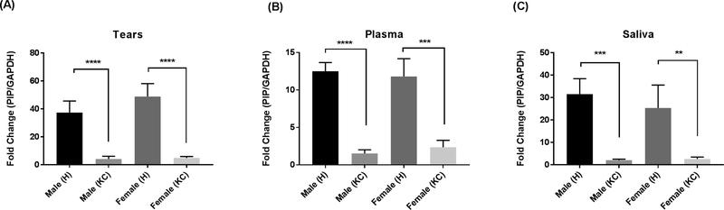
Participants: This study included one hundred and forty-seven patients with KC (105 male, 42 female), and sixty healthy controls (27 male, 33 female).

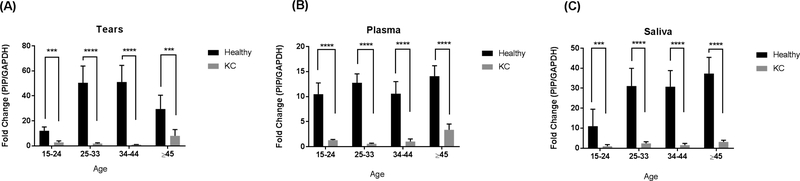
Methods: Tears, plasma and saliva samples were collected from all participants. In both KC and healthy groups all collected samples were divided into four age subgroups (15-24y), (25-34y), (35-44y) and (45y and up). Samples were analyzed using western blot (WB) and enzyme-linked immunosorbent assay (ELISA). Areas under the receiver operating characteristic curves (AUROCs) were used to evaluate diagnostic accuracy for distinguishing between KC and healthy eyes.

Main outcome measures: Difference in PIP protein levels between patients with KC and healthy controls.

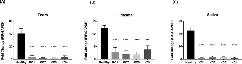
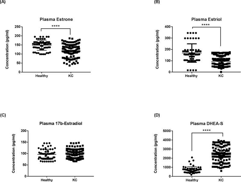
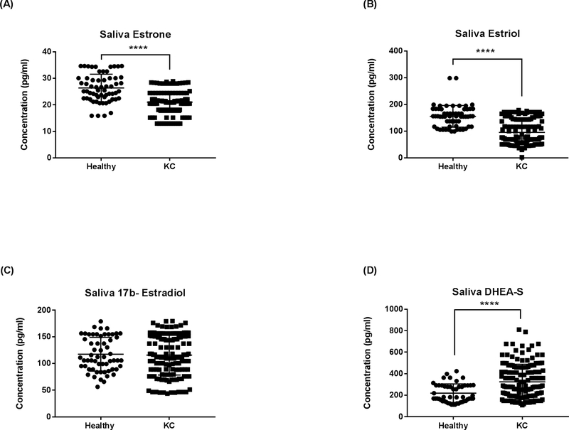
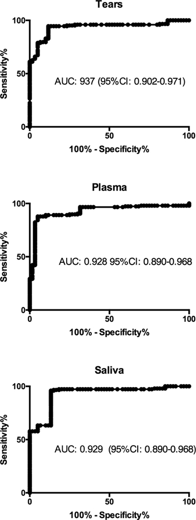
Results: Results showed significant downregulation of PIP expression in all three biological fluids on KC patients when compared to healthy controls, independent of age, sex and severity. Since PIP is a hormonal-regulated protein, we also investigated the expression of major sex hormones. We detected significant upregulation in salivary and plasma Dehydroepiandrosterone sulfate (DHEA-S) levels and significant downregulation of estrone and estriol levels, in KC patients compared to healthy controls, independent of sex, age, and KC severity stage. ROC was used to determine the overall predictive accuracy of this protein in KC. Data showed an area under the curve (AUC) for PIP in tears of 0.937 (95%CI: 0.902-0.971), in plasma of 0.928 (95%CI: 0.890-0.968) and in saliva of 0.929 (95%CI: 0.890-0.968).

Conclusions: Conclusively, our results show that PIP levels are reduced in all three human biological fluids tested, and may independently or in combination with current imaging techniques aid in screening and diagnosis of KC. Our data revealed that PIP levels can potentially differentiate between disease and healthy cases, and PIP levels are stable in relation to KC severity, sex and age. Moreover, alterations in sex hormone levels in correlation with reduced PIP levels in KC provide an intriguing insight in the underlying KC pathophysiology and highlights the role of PIP as a KC biomarker.

Keywords: Biomarker; Blood; Keratoconus; Prolactin-Induced Protein; Saliva; Tears.


Conflict of interest statement

Declarations of interest: no conflicting relationship exists for any author.

Figures

Fig. 1.
Fig. 1.
PIP Protein levels measured by western blot analysis from three biological fluids: A: Tears; B: Plasma; and C: Saliva, in patients with KC (n = 147) compared to healthy controls (n = 60). Estimates represent mean and error bars represent standard error of the mean. Mann-Whitney U test was used to test group difference, ***p < 0.001, ****p < 0.0001.
Fig. 2.
Fig. 2.
PIP Protein levels measured by western blot analysis from three biological fluids: A: Tears; B: Plasma; and C: Saliva, in patients with KC (n = 147) compared to healthy controls (n = 60). Four different age groups (15–14 yr, 25–33 yr, 34–44 yr, ≥45 yr) were analyzed and compared. Estimates represent mean and error bars represent standard error of the mean. Mann-Whitney U test was used to test group difference, ***p < 0.001, ****p < 0.0001.
Fig. 3.
Fig. 3.
PIP Protein levels measured by western blot analysis from three biological fluids: A: Tears; B: Plasma; and C: Saliva, in patients with KC (n = 147) compared to healthy controls (n = 60). Male (H): healthy male: n = 27; Male (KC): male with KC: n = 105; Female (H): healthy female: n = 33; and Female (KC): female with KC: n = 42. Estimates represent mean and error bars represent standard error of the mean. Mann-Whitney U test was used to test group difference, **p < 0.01, ***p < 0.001, ****p < 0.0001.
Fig. 4.
Fig. 4.
PIP Protein levels measured by western blot analysis from three biological fluids: A: tears, B: Plasma and C: Saliva in patients with KC (n = 147) compared to healthy controls (n = 60). Four KC severity stages were classified according to Kmax values (KC1, KC2, KC3, and KC4). Estimates represent mean and error bars represent standard error of the mean. Mann-Whitney U test was used to test group difference, ***p < 0.001, ****p < 0.0001.
Fig. 5.
Fig. 5.
ELISA analysis of total plasma sex hormone concentrations in KCs and healthy controls: (A) Estrone, (B) estriol, (C) 17β-estradiol, and (D) DHEA-S. Statistical significance was determined using Mann-Whitney U test with p ≤ 0.05 considered statistically significant. Estimates represent mean and error bars represent standard error of the mean. ****p < 0.0001.
Fig. 6.
Fig. 6.
ELISA analysis of total Saliva sex hormone concentrations in KCs and healthy controls: (A) Estrone, (B) estriol, (C) 17β-estradiol, and (D) DHEA-S. Statistical significance was determined using Mann-Whitney U test with p ≤ 0.05 considered statistically significant. Estimates represent mean and error bars represent standard error of the mean, ****p < 0.0001.
Fig 7:
Fig 7:
Receiver operating characteristic (ROC) curves for KC patients compared to healthy controls for PIP levels in tears, plasma and saliva. Area under the curve (AUC) and 95%CI quantified the ability of PIP levels to discriminate between individuals with KC and healthy controls.

References

    1. Abu-Amero KK, Al-Muammar AM, Kondkar AA, 2014. Genetics of Keratoconus: Where Do We Stand? Journal of Ophthalmology 2014, 11. - PMC - PubMed
    1. Baldini C, Ferro F, Elefante E, Bombardieri S, 2018. Biomarkers for Sjögren’s syndrome. Biomarkers in Medicine 12, 275–286. - PubMed
    1. Baniwal SK, Chimge N-O, Jordan VC, Tripathy D, Frenkel B, 2013. Prolactin-Induced Protein (PIP) Regulates Proliferation of Luminal A Type Breast Cancer Cells in an Estrogen-Independent Manner. PLoS ONE 8, e62361. - PMC - PubMed
    1. Baniwal SK, Little GH, Chimge NO, Frenkel B, 2012. Runx2 controls a feed-forward loop between androgen and prolactin-induced protein (PIP) in stimulating T47D cell proliferation. Journal of cellular physiology 227, 2276–2282. - PMC - PubMed
    1. Carsol JL, Gingras S, Simard J, 2002. Synergistic action of prolactin (PRL) and androgen on PRL-inducible protein gene expression in human breast cancer cells: a unique model for functional cooperation between signal transducer and activator of transcription-5 and androgen receptor. Mol Endocrinol 16, 1696–1710. - PubMed

Publication types

MeSH terms