. 2018 Nov 5:24:177-187.
doi: 10.12659/MSMBR.910522.

MicroRNA-32 Regulates Development and Progression of Hepatocellular Carcinoma by Targeting ADAMTS9 and Affects Its Prognosis

Affiliations

MicroRNA-32 Regulates Development and Progression of Hepatocellular Carcinoma by Targeting ADAMTS9 and Affects Its Prognosis

Shengmian Li et al. Med Sci Monit Basic Res. .

Abstract

BACKGROUND MicroRNA-32 (miR-32) induces cell proliferation and metastasis in hepatocellular carcinoma (HCC), but the detailed mechanisms of miR-32 in regulating oncogenesis and development of HCC have not been clarified. The aim of this study was to investigate the effects of miR-32 on HCC and its clinical pathological significance, as well as to determine the functional connection between miR-32 and ADAMTS9 in HCC. MATERIAL AND METHODS Quantitative RT-PCR was used to assess the expression levels of miR-32 in HCC tissues, adjacent non-cancerous tissues, and liver cancer cell lines. In vitro cell proliferation, migration, and invasion assays were performed to confirm the biological functions of miR-32. Quantitative RT-PCR, Western blot analysis, and luciferase reporter assays were used to evaluate the role of miR-32 in the regulation of ADAMTS9. RESULTS miR-32 was highly expressed in HCC tissues compared with corresponding adjacent non-cancerous tissues. Over-expression of miR-32 was also found in 3 human liver cancer cell lines: SMMC-7721, Huh7, and HepG2. Moreover, increasing expression of miR-32 in HCC tissues was related to shorter overall survival. In vitro over-expression of miR-32 promoted cell proliferation, migration, and invasion; however, the under-expression of miR-32 revealed the opposite effects. Dual-luciferase reporter assay indicated that miR-32 can directly bind to the 3'-UTR of ADAMTS9. Western blot analysis showed that over-expression of miR-32 decreased expression of ADAMTS9 protein. Rescue tests further verified the connection between miR-32 and ADAMTS9. CONCLUSIONS Our data indicate that miR-32 accelerates progression in HCC by targeting ADAMTS9, and the abnormal expression of miR-32 is correlated with prognosis and could become a potential therapeutic target.


Conflict of interest statement

Conflict of interest:

None.

Figures

Figure 1
Figure 1
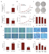
Expression of miR-32 in HCC tissues and cell lines and correlation between its expression and prognosis. (A) Expression level of miR-32 in HCC tissues was significantly increased compared to corresponding adjacent non-cancerous tissues. (B) Expression of miR-32 was markedly increased in 3 human liver cancer cell lines – SMMC-7721, Huh7, and HepG2 – and no statistically significant difference was found among these cell lines. (C) Survival curve for overall survival of HCC patients referring to different expressions of miR-32. Increasing expression of miR-32 in HCC tissues was significantly related to poor outcome.
Figure 2
Figure 2
Effects of miR-32 on biological behaviors of Huh7 cells. (A) Increased miR – 32 expression in Huh7 cells transfected with miR – 32-mimics and decreased miR – 32 expression in Huh7 cells transfected with miR-32-inhibitor were confirmed by qRT-PCR. (B) MTS assay showed that miR-32 strongly stimulated cell proliferation, while cell proliferation was significantly impaired in cells transfected with miR-32 inhibitor compared with controls. (C) Clonogenic assay validated effect of miR-32 on cell proliferation. (D) Migration and invasion ability of cells in miR32-mimics group was significantly higher as measured by wound healing assay, while in the miR32inhibitor group it was less than that of the negative group. (E) Transwell assay showed migration and invasion in Huh7 cells transfected with miR – 32-mimics and in cells transfected with miR-32-inhibitor.
Figure 3
Figure 3
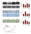
Effect of ADAMTS9 on growth and migration in HCC cells. (A) The miR-32 binding site in ADAMTS9 mRNA 3′-UTR predicted by TargetScan. (B) The relative expression of ADAMTS9 mRNA was significantly down-regulated in miR-32 transfected cells compared with the miR-32-NC group, whereas it was up-regulated in the miR-32-inhibitor transfected group. (C) Western blotting showed that expression of ADAMTS9 protein level was increased in HCC cells transfected with miR-32- inhibitor. (D) Immunohistochemical detection of ADAMTS9 expression in hepatocellular carcinoma. Appearance of granular stain in the cytoplasm was defined as positive expression. (E) Western blotting showed expression of ADAMTS9 protein was significantly increased in cells transfected with p-cDNA3.1-ADAMTS9. (F) Proliferation was inhibited in Huh7 cells transfected with p-cDNA3.1-ADAMTS9 as measured by MTS assay. (G) Cell migration and invasion were suppressed in Huh7 cells transfected with p-cDNA3.1-ADAMTS9 as determined by wound healing assay. (H) Transwell assay demonstrated that over-expression of ADAMTS9 significantly suppressed the invasion of Huh7 cells.
Figure 4
Figure 4
Effect of ADAMTS9 to biological effects of miR-32. (A) Expression of ADAMTS9 in Huh7 cells co-transfected with miR-32 and ADAMTS9 was significantly increased compared with cells only transfected with miR-32. (B, C) Wound healing and Transwell assays revealed that migration and invasion ability of Huh7 cells co-transfected with miR-32 and ADAMTS9 decreased compared with cells only transfected with miR-32, and was increasing compared with cells only transfected with ADAMTS9. (D) MTS assay confirmed that cell proliferation in Huh7 cells co-transfected with miR-32 and ADAMTS9 was inhibited compared with cells only transfected with miR-32, but it was increased in cells only transfected with ADAMTS9.
Figure 5
Figure 5
miR-32 targeted 3′-UTR of ADAMTS9. The relative luciferase activity in the PmiR-ADAMTS9-wt group transfected with miR-32 and transfected with miR-32-NC.

Similar articles

Cited by

References

    1. Farinati F, Vitale A, Spolverato G, et al. Development and validation of a new prognostic system for patients with hepatocellular carcinoma. PLoS Med. 2016;13:e1002006. - PMC - PubMed
    1. Omata M, Cheng AL, Kokudo N, et al. Asia–Pacific clinical practice guidelines on the management of hepatocellular carcinoma: A 2017 update. Hepatol Int. 2017;11:317–70. - PMC - PubMed
    1. Liu LL, Lu SX, Li Min, et al. FoxD3-regulated microRNA-137 suppresses tumor growth and metastasis in human hepatocellular carcinoma by targeting AKT2. Oncotarget. 2014;13:5113–24. - PMC - PubMed
    1. Zhang JF, Kuai XL, Song MJ, et al. MicroRNA 32 inhibits the proliferation and invasion of the SGC 7901 gastric cancer cell line in vitro. Oncol Lett. 2014;7:270–74. - PMC - PubMed
    1. Cai LZ, Cai X. Up-regulation of miR-9 expression predicate advanced clinicopathological features and poor prognosis in patients with hepatocellular carcinoma. Diagn Pathol. 2014;9:1000. - PMC - PubMed

MeSH terms