

Characteristics of surfactant proteins in tumorigenic and inflammatory lung lesions in rodents
- PMID: 30393427
- PMCID: PMC6206284
- DOI: 10.1293/tox.2018-0025
Characteristics of surfactant proteins in tumorigenic and inflammatory lung lesions in rodents
Abstract
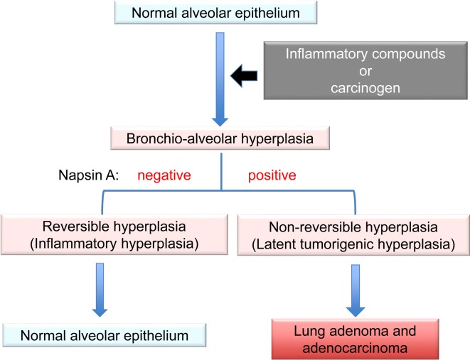
Surfactant proteins (SPs) are essential for the proper structure and respiratory function of the lungs. There are four subtypes of SPs: SP-A, SP-B, SP-C, and SP-D. The expectorant drug ambroxol hydrochloride is clinically used to stimulate pulmonary surfactant and airway serous secretion. In addition, previous studies showed that ambroxol regulated SP production and attenuated pulmonary inflammation, with ambroxol hydrochloride being found to suppress quartz-induced lung inflammation via stimulation of pulmonary surfactant and airway serous secretion. In this study, we investigated the expression of SP-A, SP-B, SP-C, and SP-D in neoplastic and inflammatory lung lesions in rodents, as well as their possible application as potential markers for diagnostic purposes. SP-B and SP-C showed strong expression in lung hyperplasia and adenoma, whereas SP-A and SP-D were expressed in the mucus or exudates of inflammatory alveoli. Rodent tumorigenic hyperplasic tissues induced by various carcinogens were positive for napsin A, an aspartic proteinase involved in the maturation of SP-B; this indicated a focal increase in type II pneumocytes in the lungs. Therefore, high expression of napsin A in the alveolar walls may serve as a useful marker for prediction of the tumorigenic potential of lung hyperplasia in rodents.
Keywords: ambroxol hydrochloride; hyperplasia; lungs; napsin A; surfactant protein.
Figures
Similar articles
-
Suppressive effects of the expectorant drug ambroxol hydrochloride on quartz-induced lung inflammation in F344 rats.J Toxicol Pathol. 2017 Apr;30(2):153-159. doi: 10.1293/tox.2016-0050. Epub 2016 Dec 22. J Toxicol Pathol. 2017. PMID: 28458453 Free PMC article.
-
Validating the use of napsin A as a marker for identifying tumorigenic potential of lung bronchiolo-alveolar hyperplasia in rodents.Exp Toxicol Pathol. 2017 Oct 2;69(8):637-642. doi: 10.1016/j.etp.2017.06.001. Epub 2017 Jun 8. Exp Toxicol Pathol. 2017. PMID: 28602391
-
Immunohistochemical characteristics of surfactant proteins a, B, C and d in inflammatory and tumorigenic lung lesions of f344 rats.J Toxicol Pathol. 2014 Oct;27(3-4):175-82. doi: 10.1293/tox.2014-0020. Epub 2014 Jun 9. J Toxicol Pathol. 2014. PMID: 25378802 Free PMC article.
-
Molecular and cellular processing of lung surfactant.FASEB J. 1994 Sep;8(12):957-67. doi: 10.1096/fasebj.8.12.8088461. FASEB J. 1994. PMID: 8088461 Review.
-
Hormonal and developmental regulation of pulmonary surfactant synthesis in fetal lung.Baillieres Clin Endocrinol Metab. 1990 Jun;4(2):351-78. doi: 10.1016/s0950-351x(05)80055-2. Baillieres Clin Endocrinol Metab. 1990. PMID: 2248600 Review.
Cited by
-
Single Intratracheal Quartz Instillation Induced Chronic Inflammation and Tumourigenesis in Rat Lungs.Sci Rep. 2020 Apr 20;10(1):6647. doi: 10.1038/s41598-020-63667-4. Sci Rep. 2020. PMID: 32313071 Free PMC article.
-
Relationship between Lung Carcinogenesis and Chronic Inflammation in Rodents.Cancers (Basel). 2021 Jun 10;13(12):2910. doi: 10.3390/cancers13122910. Cancers (Basel). 2021. PMID: 34200786 Free PMC article. Review.
References
-
- Pérez-Gil J, and Keough KM. Interfacial properties of surfactant proteins. Biochim Biophys Acta. 1408: 203–217. 1998. - PubMed
-
- Perez-Gil J, and Weaver TE. Pulmonary surfactant pathophysiology: current models and open questions. Physiology (Bethesda). 25: 132–141. 2010. - PubMed
-
- Yokohira M, Yamakawa K, Nakano Y, Numano T, Furukawa F, Kishi S, Ninomiya F, Kanie S, Hitotsumachi H, Saoo K, and Imaida K. Immunohistochemical characteristics of surfactant proteins a, B, C and d in inflammatory and tumorigenic lung lesions of f344 rats. J Toxicol Pathol. 27: 175–182. 2014. - PMC - PubMed
-
- Houtmeyers E, Gosselink R, Gayan-Ramirez G, and Decramer M. Effects of drugs on mucus clearance. Eur Respir J. 14: 452–467. 1999. - PubMed
Publication types
LinkOut - more resources
Full Text Sources