. 2018 Jul 30;7(9):e1468954.
doi: 10.1080/2162402X.2018.1468954. eCollection 2018.

Wiskott-Aldrich syndrome gene mutations modulate cancer susceptibility in the p53± murine model

Affiliations

Wiskott-Aldrich syndrome gene mutations modulate cancer susceptibility in the p53± murine model

Marton Keszei et al. Oncoimmunology. .

Abstract

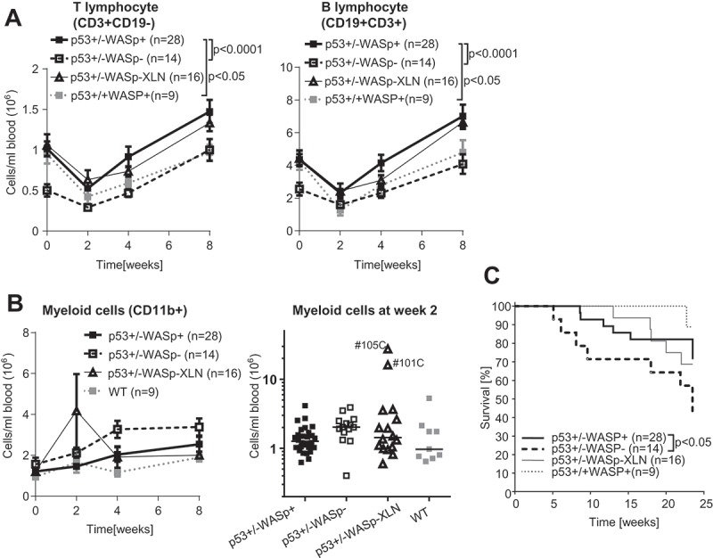
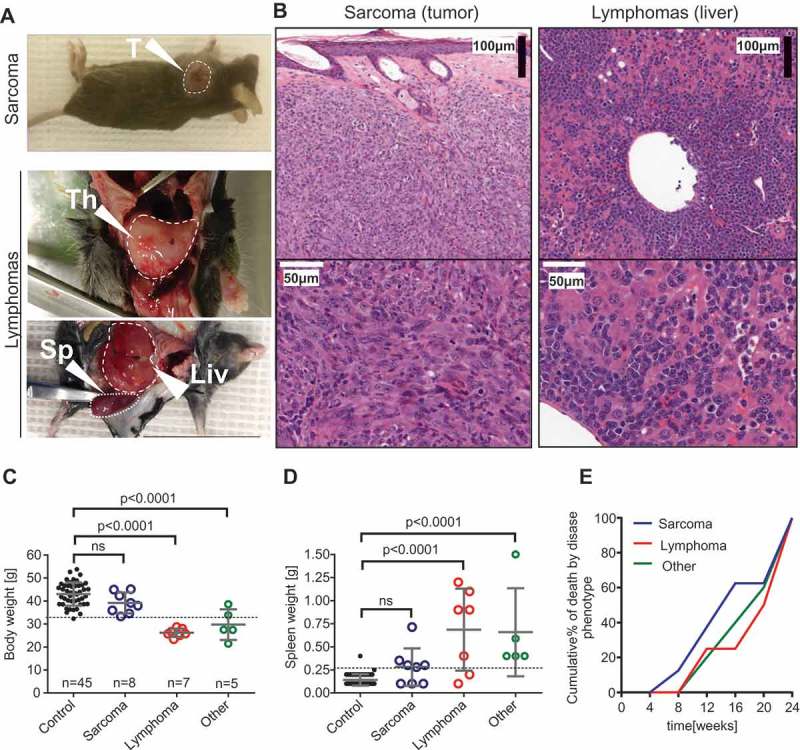
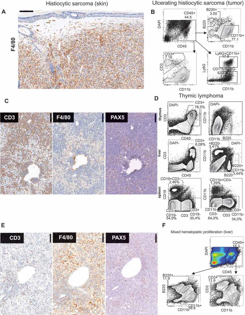
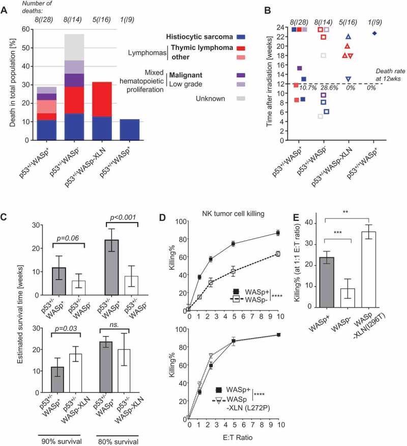
The Wiskott-Aldrich syndrome protein (WASp) is a key regulator of the actin cytoskeleton in hematopoietic cells and mutated in two severe immunodeficiency diseases with high incidence of cancer. Wiskott-Aldrich syndrome (WAS) is caused by loss-of-function mutations in WASp and most frequently associated with lymphoreticular tumors of poor prognosis. X-linked neuropenia (XLN) is caused by gain-of-function mutations in WASp and associated with acute myeloid leukemia (AML) and myelodysplastic syndrome (MDS). To understand the role of WASp in tumorigenesis, we bred WASp+, WASp-, and WASp-XLN mice onto tumor susceptible p53+/- background and sub-lethally irradiated them to enhance tumor development. We followed the cohorts for 24 weeks and tumors were characterized by histology and flow cytometry to define the tumor incidence, onset, and cell origin. We found that p53+/-WASp+ mice developed malignancies, including solid tumors and T cell lymphomas with 71.4% of survival 24 weeks after irradiation. p53+/-WASp- mice showed lower survival rate and developed various early onset malignancies. Surprisingly, the p53+/-WASp-XLN mice developed malignancy mostly with late onset, which caused delayed mortality in this colony. This study provides evidence for that loss-of-function and gain-of-function mutations in WASp influence tumor incidence and onset.

Keywords: WASp; genetic model; immunodeficiency; malignancies; p53.


Figures

Figure 1.
Figure 1.
Blood leucocyte counts and survival rate in the p53+/-WASp mutant tumor model. Blood lymphocyte (A) and (B) myeloid cell counts were determined with flow cytometry for 8 weeks after irradiation. Two-way ANOVA and Bonferroni’s multiple comparison test. Mean±SEM. (B right panel: median myeloid count) (C) Kaplan-Maier survival curve of p53+/-WASp+, p53+/-WASp, p53+/-WASp XLN, and p53+/+WASp+ mice. (p value: Gehan-Breslow-Wilcoxon test).
Figure 2.
Figure 2.
Phenotypical features of malignancies in the p53+/- irradiation model. (A) Sarcomas (upper panel) were initially identified as visible tumors on exteriors. Lymphomas (lower panels) were recognized in animals showing severe weight loss, enlarged thymus or a combination of extreme splenomegaly and other microscopic features. T: tumor; Th: thymus; Sp: spleen; Liv: liver. (B) Hematoxylin-eosin stained tissue sections from sarcoma (#135A left panel) and lymphoma (#65B right panel). (C) Body weight before sacrificing in control (survived until endpoint) mice vs. mice with various pathology and (D) spleen weight at endpoint in control (survived until endpoint) mice vs. mice with various pathology. ANOVA and Bonferroni’s multiple comparison test. Mean±SD. (E) Cumulative % of tumor deaths was calculated by binning the frequencies of deaths into 4 weeks periods.
Figure 3.
Figure 3.
Immuno phenotypical features of maligancies in p53+/- irradiation model. (A) F4/80 immunohistochemistry of subcutaneous tumor. (#135A) (B) FACS analysis of large ulcerating tumor. (#87A) (C) Immunohistochemistry of liver in lymphoma with the indicated antibodies. (#124A). (D) FACS analysis of thymus, liver, and spleen in mouse with thymic lymphoma. (#78A) (E) Immunohistochemistry of liver of mouse with severe mixed hematopoietic proliferation. (#44C). (F) FACS Liver (#114A).
Figure 4.
Figure 4.
Frequency of deaths in p53+/-WASp mutant tumor model. (A) Frequency of deaths per genotype. Bold text indicates malignant cause of death. (B) Time of death after irradiation. Causes of death are color coded as on Figure 4A. (C) Survival time and 95% confidence intervals at 10th and 20th survival percentiles was calculated from the Figure 1C survival curve with Laplace regression. (D, E) Killing of YAC-1-luciferase tumor cells by NK cells was quantified with chemiluminescence. Mean±SD; Two-way ANOVA (D) and ANOVA with Bonferroni’s multiple comparisons test (E). **: p ≤ 0.01, ***: p ≤ 0.001, ****: p ≤ 0.0001.

Similar articles

Cited by

References

    1. Mortaz E, Tabarsi P, Mansouri D, Khosravi A, Garssen J, Velayati A, Adcock IM.. Cancers related to immunodeficiencies: update and perspectives. Front Immunol. 2016;7:365. doi:10.3389/fimmu.2016.00365. - DOI - PMC - PubMed
    1. Imai K, Morio T, Zhu Y, Jin Y, Itoh S, Kajiwara M, Yata J, Mizutani S, Ochs HD, Nonoyama S. Clinical course of patients with WASP gene mutations. Blood. 2004;103:456–464. doi:10.1182/blood-2003-05-1480. - DOI - PubMed
    1. Sullivan KE, Mullen CA, Blaese RM, Winkelstein JA. A multiinstitutional survey of the Wiskott-Aldrich syndrome. J Pediatr. 1994;125:876–885. doi:10.1016/S0022-3476(05)82002-5. - DOI - PubMed
    1. Tran H, Nourse J, Hall S, Green M, Griffiths L, Gandhi MK. Immunodeficiency-associated lymphomas. Blood Rev. 2008;22:261–281. doi:10.1016/j.blre.2008.03.009. - DOI - PubMed
    1. Senapati J, Devasia AJ, David S, Manipadam MT, Nair S, Jayandharan GR, George B. Diffuse large B cell lymphoma in wiskott-Aldrich syndrome: a case report and review of literature. Indian J Hematol Blood Transfus. 2014;30:309–313. doi:10.1007/s12288-014-0377-1. - DOI - PMC - PubMed

Publication types