

Optimized Diffusion-Weighting Gradient Waveform Design (ODGD) formulation for motion compensation and concomitant gradient nulling
- PMID: 30394568
- PMCID: PMC6289642
- DOI: 10.1002/mrm.27462
Optimized Diffusion-Weighting Gradient Waveform Design (ODGD) formulation for motion compensation and concomitant gradient nulling
Abstract
Purpose: To present a novel Optimized Diffusion-weighting Gradient waveform Design (ODGD) method for the design of minimum echo time (TE), bulk motion-compensated, and concomitant gradient (CG)-nulling waveforms for diffusion MRI.
Methods: ODGD motion-compensated waveforms were designed for various moment-nullings Mn (n = 0, 1, 2), for a range of b-values, and spatial resolutions, both without (ODGD-Mn ) and with CG-nulling (ODGD-Mn -CG). Phantom and in-vivo (brain and liver) experiments were conducted with various ODGD waveforms to compare motion robustness, signal-to-noise ratio (SNR), and apparent diffusion coefficient (ADC) maps with state-of-the-art waveforms.
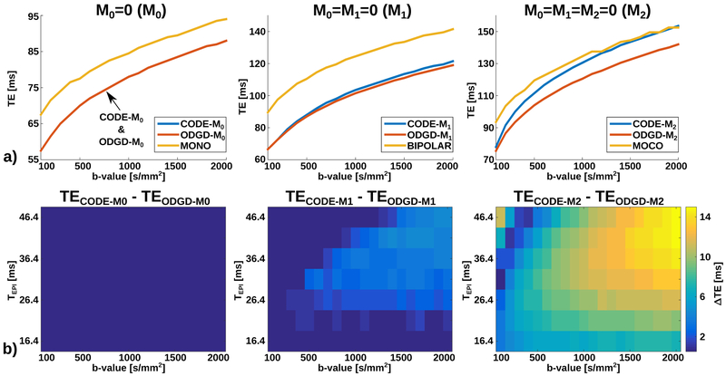
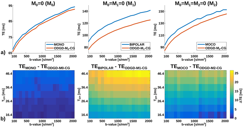
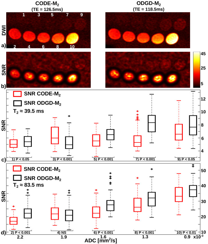
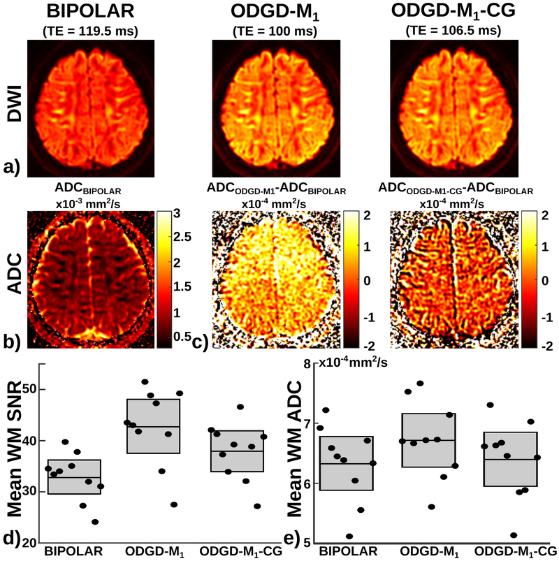
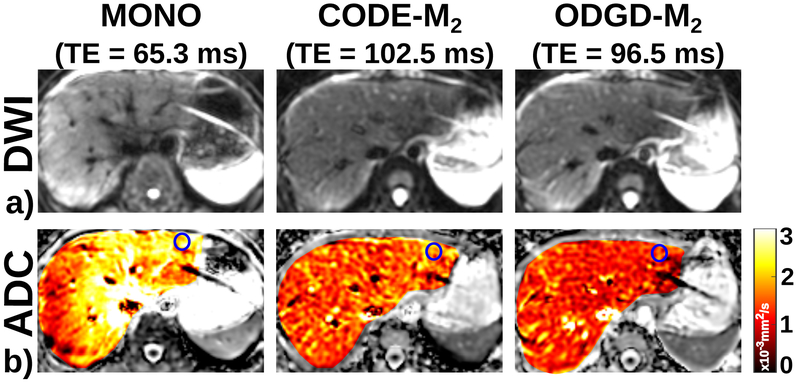
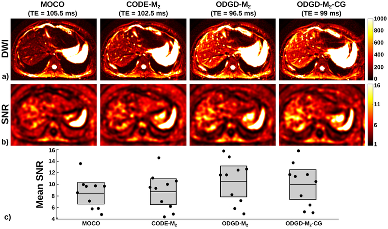
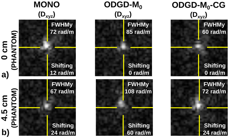
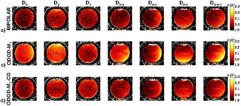
Results: ODGD-Mn and ODGD-Mn -CG waveforms reduced the TE of state-of-the-art waveforms. This TE reduction resulted in significantly higher SNR (P < 0.05) in both phantom and in-vivo experiments. ODGD-M1 improved the SNR of BIPOLAR (42.8 ± 5.3 vs. 32.9 ± 3.3) in the brain, and ODGD-M2 the SNR of motion-compensated (MOCO) and Convex Optimized Diffusion Encoding-M2 (CODE-M2 ) (12.3 ± 3.6 vs. 9.7 ± 2.9 and 10.2 ± 3.4, respectively) in the liver. Further, ODGD-M2 also showed excellent motion robustness in the liver. ODGD-Mn -CG waveforms reduced the CG-related dephasing effects of non CG-nulling waveforms in phantom and in-vivo experiments, resulting in accurate ADC maps.
Conclusions: ODGD waveforms enable motion-robust diffusion MRI with reduced TEs, increased SNR, and reduced ADC bias compared to state-of-the-art waveforms in theoretical results, simulations, phantoms and in-vivo experiments.
Keywords: Concomitant Gradient (CG)-nulling; Diffusion-Weighted Imaging (DWI); diffusion-weighting gradient waveforms; motion compensation; optimization.
© 2018 International Society for Magnetic Resonance in Medicine.
Figures

References
-
- Taouli B, Koh DM. Diffusion-weighted MR imaging of the liver 1. Radiology 2009;254:47–66. - PubMed
-
- Koh DM, Collins DJ. Diffusion-weighted MRI in the body: applications and challenges in oncology. American Journal of Roentgenology 2007;188:1622–1635. - PubMed
-
- Stejskal EO, Tanner JE. Spin diffusion measurements: spin echoes in the presence of a time-dependent field gradient. The journal of chemical physics 1965;42:288–292.
-
- Le Bihan D, Breton E, Lallemand D, Grenier P, Cabanis E, Laval-Jeantet M. MR imaging of intravoxel incoherent motions: application to diffusion and perfusion in neurologic disorders. Radiology 1986;161:401–407. - PubMed
MeSH terms
Substances
Grants and funding
LinkOut - more resources
Full Text Sources
Other Literature Sources
