Comparative Study
. 2019 Jul 1;115(8):1332-1342.
doi: 10.1093/cvr/cvy278.

Protein kinase C binding protein 1 inhibits hypoxia-inducible factor-1 in the heart

Affiliations
Comparative Study

Protein kinase C binding protein 1 inhibits hypoxia-inducible factor-1 in the heart

Kathryn J Schunke et al. Cardiovasc Res. .

Abstract

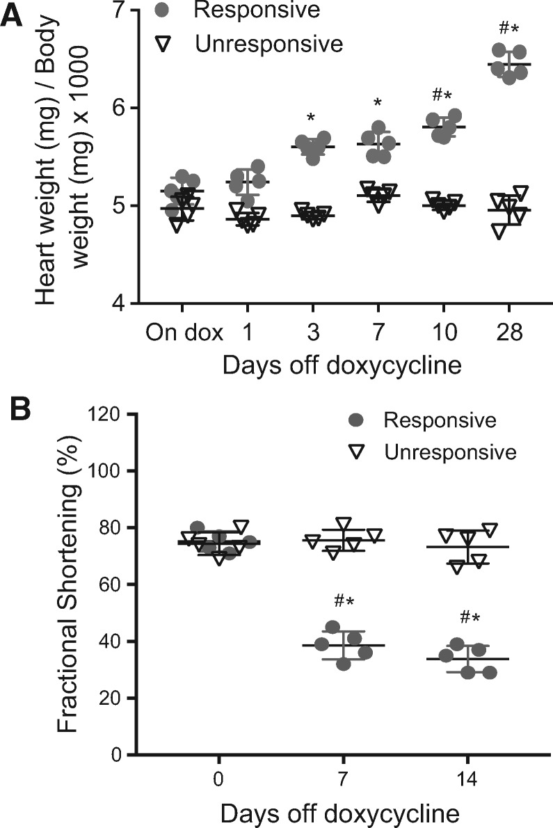
Aims: Hypoxia-inducible factor-1 alpha (HIF-1α) is a key transcription factor responsible for the induction of genes that facilitate adaptation to hypoxia. To study HIF-1 signalling in the heart, we developed a mouse model in which an oxygen-stable form of HIF-1α can be inducibly expressed in cardiac myocytes, under the regulation of tetracycline.

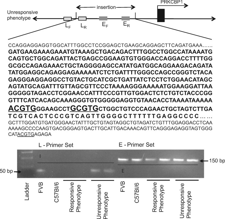
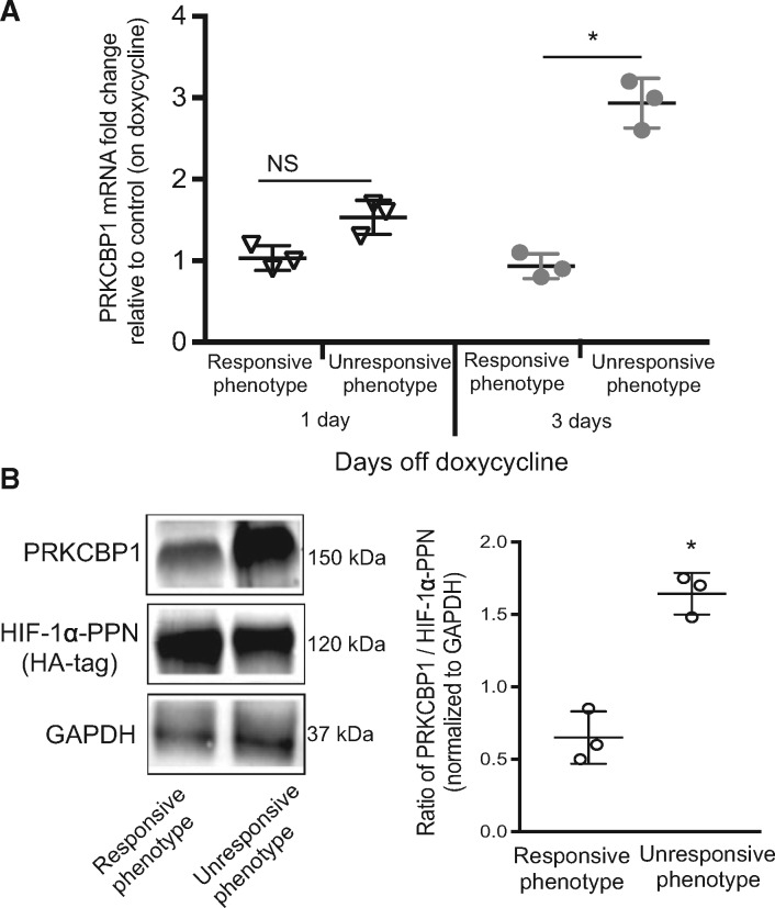
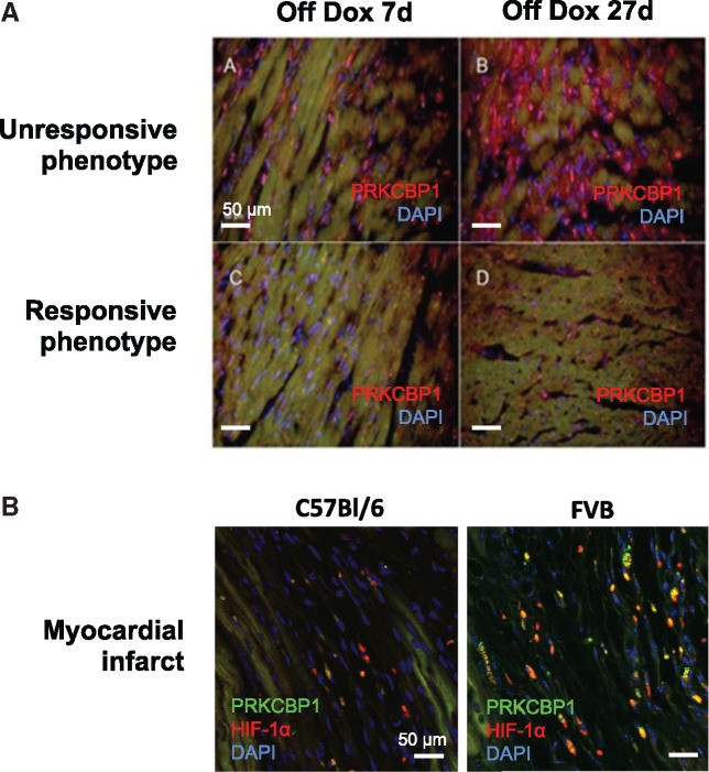
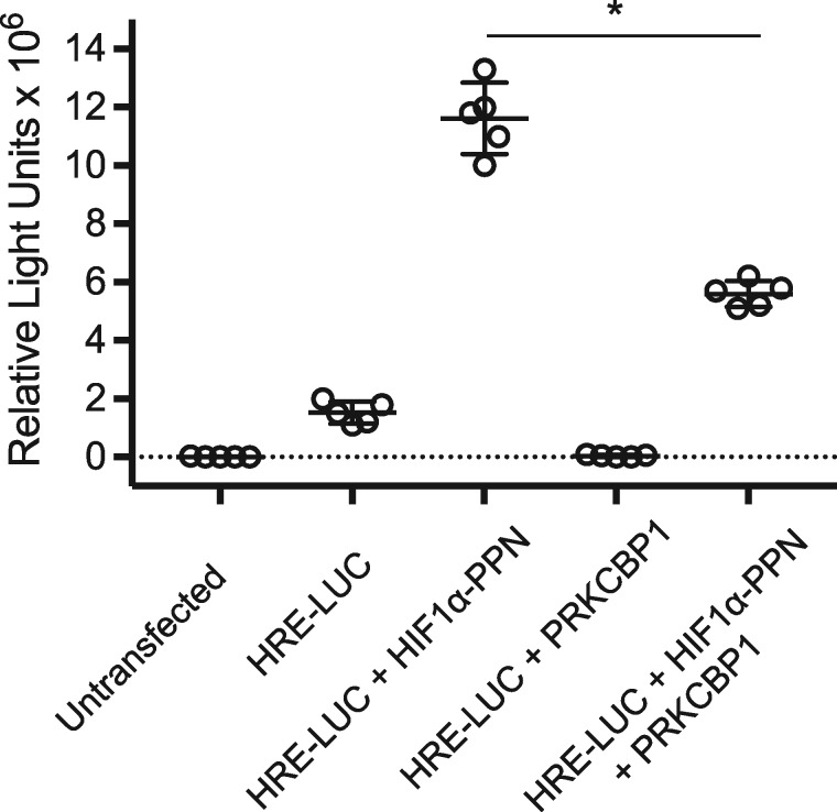
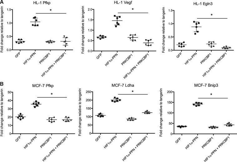
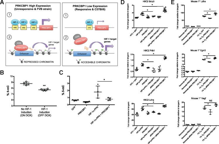
Methods and results: Remarkably, expression of the transgene in mice generated two distinct phenotypes. One was the expected expression of HIF-regulated transcripts and associated changes in cardiac angiogenesis and contractility. The other was an unresponsive phenotype with much less expression of typical HIF-response genes and substantial expression of a zinc-finger protein, Protein Kinase C Binding Protein 1 (PRKCBP1). We have demonstrated that this second phenotype is due to an insertion of a fragment of DNA upstream of the PRKCBP1 gene that contains two additional canonical HIF binding sites and leads to substantial HIF binding, assessed by chromatin immunoprecipitation, and transcriptional activation. This insertion is found only in the FVB strain of mice that contributed the αMHC-tet binding protein transgene to these biallelic mice. In HEK293 cells transfected with oxygen-stable HIF-1α and PRKCBP1, we demonstrated inhibition of HIF-1 activity by a luciferase reporter assay. Using mouse primary cells and cell lines, we show that transfection with oxygen-stable HIF-1α and PRKCBP1 reduced expression of direct HIF-1 gene targets and that knockdown of PRKCBP1 removes that negative inhibition. Consistent with previous reports suggesting that PRKCBP1 modulates the chromatin landscape, we found that HL-1 cells transfected with oxygen-stable HIF-1α and PRKCBP1 have reduced global 5-methyl cytosine compared to HIF-1 alone.

Conclusion: We show genetic, transcriptional, biochemical, and physiological evidence that PRKCBP1 inhibits HIF activity. Identification of a new oxygen-dependent and previously unsuspected regulator of HIF may provide a target for new therapeutic approaches to ischaemic heart disease.

Keywords: Genetic variance; HIF inhibition; RACK7; Zmynd8; transcriptional repressor.


Figures

Figure 1
Figure 1
HIF-1α-PPN transgene phenotypes. When induced to express the HIF-1α-PPN transgene by removal of doxycycline from the drinking water, some mice do not exhibit the expected phenotype, (A) as assessed by change in heart weight compared to body weight (n = 5 per group and time point), and (B) change in ventricular function (fractional shortening %; n = 5 per group and time point). Two-way ANOVA (A) and repeated measures two-way ANOVA (B) with Tukey’s post hoc test were used for statistical analysis. Data are expressed as mean ± SD; *#P < 0.05 (*between groups and #to baseline).
Figure 2
Figure 2
Genomic ‘FVB allele’ insertion. Illustration of genomic insertion and PCR identification of variant ‘FVB allele’. The specific insertion in the Prkcbp1 gene is only found in the FVB strain and unresponsive phenotype. The 480 bp inserted sequence is shown in bold and putative HREs are underlined.
Figure 3
Figure 3
PRKCBP1 is more abundant in the unresponsive hearts. (A) Real-time PCR analysis of PRKCBP1 transcript. Hearts taken from the unresponsive mice have greater PRKCBP1 transcript than the hearts from responsive mice, assessed after 3 days of transgene induction. (B) HIF-1α and PRKCBP1 western blots and densitometry. Unresponsive hearts express significantly more PRKCBP1 with similar induction of HIF-1 transgene. HA-tag is used to assess HIF-1α-PPN expression. To reduce non-specific binding of primary antibodies, PVDF membranes were cut into three fractions, one containing the region 260–130 kDa for PRKCBP1 immunoblotting, 130–60 kDa for HIF-1α, and 60–8 kDa for GAPDH, according to pre-stained protein ladder. Two-tailed Student’s t-test was used for statistical analysis. Data are expressed as mean ± SD; n = 3 per group. *P < 0.05.
Figure 4
Figure 4
Immunohistological evaluation of PRKCBP1. (A) PRKCBP1 expression after transgene expression. Unresponsive mice express more PRKCBP1 than responsive mice at 7 days and 27 days of HIF-1α transgene induction (red: PRKCBP1 and blue: nuclear DAPI). (B) PRKCBP1 expression after myocardial infarct. Three days after induced myocardial infarct, the parental FVB strain expresses more PRKCBP1 than C57Bl/6 mice in the infarct region. (red: HIF-1α, green: PRKCBP1, and blue: nuclear DAPI).
Figure 5
Figure 5
HIF-1 activity by luciferase reporter. HIF-1 regulated luciferase activity is reduced by co-transfection of HEK293 cells with CMV-driven expression vectors for HIF-1α-PPN and PRKCBP1. Two-tailed Student’s t-test was used for statistical analysis. Data are expressed as mean ± SD; n = 5 per group. *P < 0.05. HRE-LUC, hypoxia response elements concatamerized to a luciferase reporter
Figure 6
Figure 6
In vitro co-transfection of HL-1 and MCF-7 cells. Transfection of HIF-1α-PPN leads to induction of HIF-1α target gene mRNA as assessed by semi-quantitative real-time PCR, in (A) HL-1, and (B) MCF-7. Co-transfection with a PRKCBP1 expression vector reduces HIF target gene transcript. One-way ANOVA with Bonferroni post hoc test was used for statistical analysis. n = 6. Data are expressed as mean ± SD. *P < 0.05.
Figure 7
Figure 7
Proposed mechanism. (A) Proposed mechanism of HIF-induced PRKCBP1 expression, and PRKCBP1 mediated negative feedback of HIF-1 regulated genes. (B) Global DNA methylation is altered following cardiac HIF-1 induction in mice. After 3 days of HIF-1 induction, ELISA analysis confirmed significant reduction in the number of methylated CG residues compared to HIF-1 non-induced mice. n = 4 per group, **P = 0.0007. (C) DNA methylation is altered in response to HIF-1 and PRKCBP1 in HL-1 cells. ELISA analysis confirmed significant increase in the number of methylated CG residues in HL-1 HIF-1α-PPN transfected cells, and co-transfection with PRKCBP1 reduced % 5-mC to baseline levels. n = 4 per group, *P < 0.05. PRKCBP1 modulates the HIF response in (D) H9C2 cells and (E) mouse primary cardiomyocytes. Overexpression of PRKCBP1 in the presence of (D) HIF-1α-PPN or (E) 1% O2 reduces HIF-1 target gene transcript. siRNA mediated knockdown of PRKCBP1, in the presence of HIF-1α-PPN or hypoxia removed this negative inhibition (n = 6 H9C2; n = 3 mouse primary cells). Two-tailed Student’s t-test (B) and one-way ANOVA with Bonferroni post hoc test (CE) was used for statistical analysis. Data are expressed as mean ± SD. *P < 0.05. Adapted from Shen et al. HRE, hypoxia response element; KDM, lysine demethylase.

Comment in

References

    1. Semenza GL. Hypoxia-inducible factor 1: master regulator of O2 homeostasis. Curr Opin Genet Dev 1998;8:588–594. - PubMed
    1. Shohet RV, Garcia JA.. Keeping the engine primed: HIF factors as key regulators of cardiac metabolism and angiogenesis during ischemia. J Mol Med 2007;85:1309–1315. - PubMed
    1. Wang GL, Jiang BH, Rue EA, Semenza GL.. Hypoxia-inducible factor 1 is a basic-helix-loop-helix-PAS heterodimer regulated by cellular O2 tension. Proc Natl Acad Sci USA 1995;92:5510–5514. - PMC - PubMed
    1. Masson N, Ratcliffe PJ.. HIF prolyl and asparaginyl hydroxylases in the biological response to intracellular O(2) levels. J Cell Sci 2003;116:3041–3049. - PubMed
    1. Masson N, Willam C, Maxwell PH, Pugh CW, Ratcliffe PJ.. Independent function of two destruction domains in hypoxia-inducible factor-alpha chains activated by prolyl hydroxylation. EMBO J 2001;20:5197–5206. - PMC - PubMed

Publication types

MeSH terms

Substances