Clinical Trial
. 2019 Feb;34(2):375-386.
doi: 10.1002/jbmr.3594. Epub 2018 Nov 5.

FAM92A Underlies Nonsyndromic Postaxial Polydactyly in Humans and an Abnormal Limb and Digit Skeletal Phenotype in Mice

Affiliations
Clinical Trial

FAM92A Underlies Nonsyndromic Postaxial Polydactyly in Humans and an Abnormal Limb and Digit Skeletal Phenotype in Mice

Isabelle Schrauwen et al. J Bone Miner Res. 2019 Feb.

Abstract

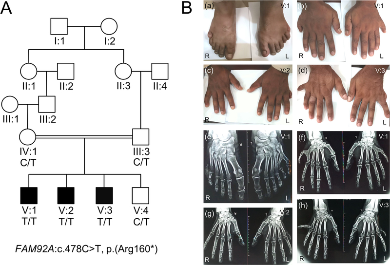
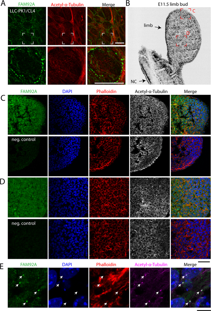
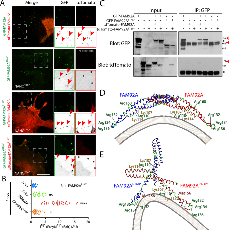
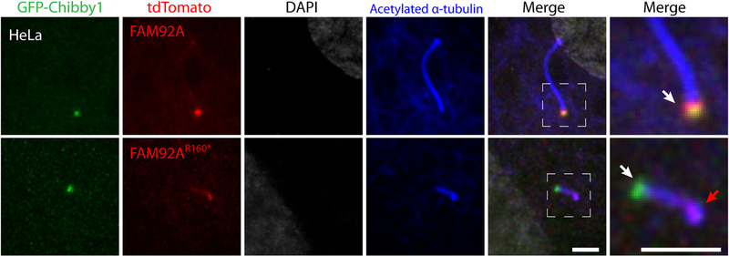
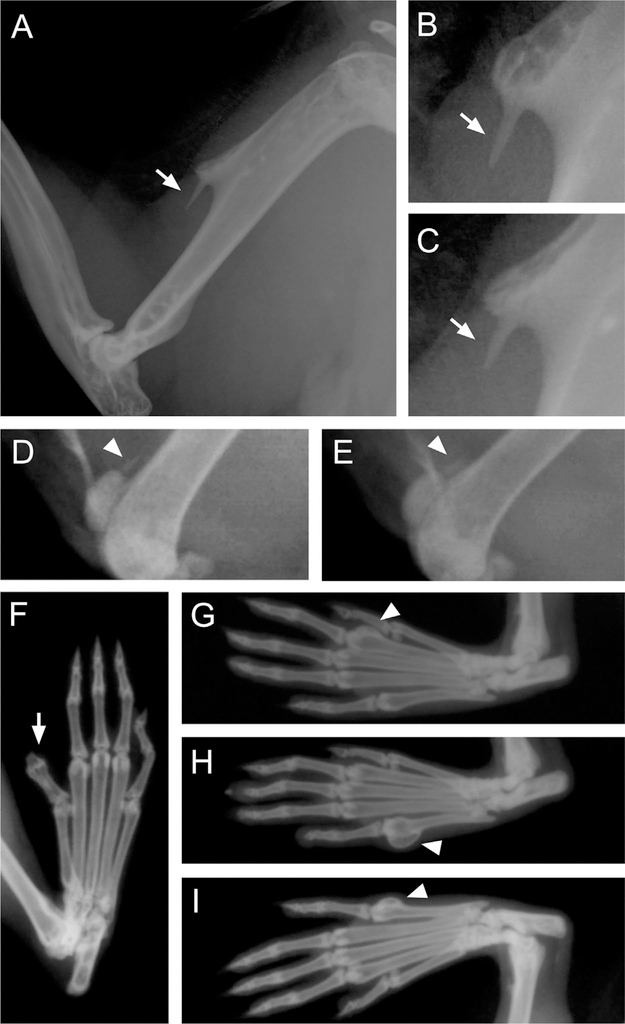
Polydactyly is a common congenital anomaly of the hand and foot. Postaxial polydactyly (PAP) is characterized by one or more posterior or postaxial digits. In a Pakistani family with autosomal recessive nonsyndromic postaxial polydactyly type A (PAPA), we performed genomewide genotyping, linkage analysis, and exome and Sanger sequencing. Exome sequencing revealed a homozygous nonsense variant (c.478C>T, p.[Arg160*]) in the FAM92A gene within the mapped region on 8q21.13-q24.12 that segregated with the PAPA phenotype. We found that FAM92A is expressed in the developing mouse limb and E11.5 limb bud including the progress zone and the apical ectodermal ridge, where it strongly localizes at the cilia level, suggesting an important role in limb patterning. The identified variant leads to a loss of the FAM92A/Chibby1 complex that is crucial for ciliogenesis and impairs the recruitment and the colocalization of FAM92A with Chibby1 at the base of the cilia. In addition, we show that Fam92a-/- homozygous mice also exhibit an abnormal digit morphology, including metatarsal osteomas and polysyndactyly, in addition to distinct abnormalities on the deltoid tuberosity of their humeri. In conclusion, we present a new nonsyndromic PAPA ciliopathy due to a loss-of-function variant in FAM92A. © 2018 American Society for Bone and Mineral Research.

Keywords: CHIBBY1; CILIOPATHY; FAM92A; PAPA; POSTAXIAL POLYDACTYLY.


Conflict of interest statement

Disclosures

All authors state that they have no conflicts of interest.

Figures

Fig. 1.
Fig. 1.
Pedigree and clinical features. (A) Pedigree drawing of family BD152 with autosomal recessive PAPA. Clear symbols represent unaffected individuals, whereas filled ones represent family members with PAPA. The individuals with a genotype had available DNA samples that underwent genomewide genotyping. The DNA sample from affected family member V:2 also underwent exome sequencing. (B) Patient V:1 showing bilateral PAPA in feet (a) and bilateral PAPA in hands (b). Patient V:2 showing postaxial extra finger in the left hand (c). Patient V:3 showing extra finger deviated to the ulnar side in the right hand and underdeveloped finger in the left hand (d). Radiograph of feet of patient V:1 showing 6th digit without metatarsal in right foot and varus deviation of the 5th and valgus deviation of the 6th toe with extra toe attached to the 5th metatarsal in left foot (e). Radiograph of hands of the same patient showing bilateral extra fingers attached to the 5th metacarpals in each hand (f). Radiograph of hands of affected individual V:2 displaying unilateral PAP in the left hand (g). Radiograph of hands of affected individual V:3 showing well-developed phalanges of 6th finger with valgus deviation in the right hand and underdeveloped phalanges of extra finger in the left hand (h). R=right; L=left.
Fig. 2.
Fig. 2.
FAM92A is strongly expressed in the embryonic mouse limb bud. (A) LLC-PK1/CL4 cells were immunostained with FAM92A (green) and acetylated alpha-tubulin (red) antibodies. (B) Inverted grayscale image of an E11.5 mouse limb bud 14 μm cryosection counterstained with phalloidin. The red boxes are showing the areas imaged in C and D. NC=notochord. (C, D) Confocal images of E11.5 mouse limb bud 14 μm cryosections immunostained with FAM92A (green) and acetylated-α-tubulin (grayscale) antibodies and counterstained with rhodamin phalloidin (red) and 4′,6-Diamidine-2′-phenylindole dihydrochloride (DAPI) (blue). The merging of green, red, and blue channels are shown. Negative control samples (neg. control) were immunostained with the secondary antibody only. (E) High magnification of cells taken from the AERof the E11.5 limb bud immunostained with FAM92A (green) and acetylated-α-tubulin (magenta) antibodies and counterstained with rhodamine phalloidin (red) and DAPI (blue). Arrowheads are pointing to the cilia. Scale bars=10 μm (A), 50 μm (C, D), and 10 μm (E).
Fig. 3.
Fig. 3.
The p.Arg160* variant disrupts FAM92A dimerization. (A) COS-7 cells were cotransfected with nonfluorescent MYO10NANOTRAP construct and GFP-FAM92AR160* (baits, green) and/or tdTomato-FAM92A or tdTomato-FAM92AR160* (Preys, red) constructs. Single channels are also shown as inverted grayscale images. Accumulations at the tip of bait and prey are shown with an arrowhead. Stars indicate the absence of accumulation of prey at the filopodia tips. Scale bar=10 μm. (B) Quantification of nanoSPD 2.0 assays. CIB2 was used as a negative control. ****p ≤ 1.0×10⁻⁴; ns=not significant. AU=Arbitrary Unit. (C) Co-immunoprecipitation assay is showing the impairment of FAM92A homodimerization by p.Arg160* variant. The red arrows indicate the expected sizes of fusion proteins. *IgG heavy chain. (D, E) 3D modeling of the quaternary structure of FAM92A/FAM92A (D) and FAM92AR160* /FAM92AR160* (E) homodimers. Arg and Lys residues are shown in green and yellow, respectively. The plasma membrane is shown in gray.
Fig. 4.
Fig. 4.
The p.Arg160* variant disrupts the FAM92A/Chibby1 complex. (A) COS-7 cells were cotransfected with nonfluorescent MYO10NANOTRAP construct, GFP-Chibby1 (bait, green), and/or tdTomato-FAM92A or tdTomato-FAM92AR160* (Preys, red) constructs. Single channels are also shown as inverted grayscale images. Accumulations at the tip of bait and prey are shown with an arrowhead. Stars indicate the absence of accumulation of prey at the filopodia tip. Scale bar=10 μm. (B) Quantification of nanoSPD 2.0 assay. CIB2 was used as a negative control. ****p ≤ 0.0001; ns=nonsignificant; AU=Arbitrary Unit. (C) Co-immunoprecipitation of FAM92A variants with Chibby1 and FAM92A variants. The red arrows indicate the expected sizes of fusion proteins. *IgG heavy chain.
Fig. 5.
Fig. 5.
The p.Arg160* variant impairs the recruitment and the colocalization of FAM92A with Chibby1 at the base of cilia. HeLa cells were cotransfected with GFP-Chibby1 (green) and tdTomato-FAM92A or tdTomato-FAM92AR160* (red) constructs. Ciliogenesis was induced by 24-hour serum starvation. HeLa cells were immunostained with acetylated α-tubulin antibody (blue), a marker of cilia, and counterstained with DAPI (grayscale). Single channel and merge images are shown. Accumulations of Chibby1 and Fam92a variants are indicated with white and red arrows. Scale bars=2 μm.
Fig. 6.
Fig. 6.
Abnormalities of the humeri and digits in homozygous Fam92a−/− mice shown by X-ray analysis. (A–C) Nine of 14 homozygous mice exhibit a similar unilateral or bilateral exostosis on the deltoid tuberosity of the humerus. This deltoid spur is compatible with a tendon calcification, particularly calcification of the m. deltoideus (scapular part). (D, E) Two Fam92a−/− mice also show tendon calcifications on their left stifle (quadriceps tendon). (F–H) Four homozygous mice showed an abnormal digit morphology. (F) Polysyndactyly of the right hind paw at phalanges. (G) Osteoma on digit two of left hind paw at the metatarsal. (H) Osteoma on digit five of the left hind paw at the metatarsal. (I) Osteoma on digit five of the right hind paw at the metatarsal. Arrows or arrowheads indicate the abnormality.

References

    1. Biesecker LG. Polydactyly: how many disorders and how many genes? 2010 update. Dev Dyn. 2011;240(5):931–42. - PMC - PubMed
    1. Castilla EE, Lugarinho R, da Graça Dutra M, Salgado LJ. Associated anomalies in individuals with polydactyly. Am J Med Genet. 1998;80(5):459–65. - PubMed
    1. Verma PK, El-Harouni AA. Review of literature: genes related to postaxial polydactyly. Front Pediatr. 2015;3:8. - PMC - PubMed
    1. Galjaard R-JH, Smits APT, Tuerlings JHAM, et al. A new locus for postaxial polydactyly type A/Bon chromosome 7q21-q34. Eur J Hum Genet. 2003;11(5):409–15. - PubMed
    1. Zhao H, Tian Y, Breedveld G, et al. Postaxial polydactyly type A/B (PAP-A/B) is linked to chromosome 19p13.1–13.2 in a Chinese kindred. Eur J Hum Genet. 2002;10(3):162–6. - PubMed

Publication types

MeSH terms

Supplementary concepts