. 2018 Dec 3;128(12):5489-5504.
doi: 10.1172/JCI99629. Epub 2018 Nov 5.

Biallelic mutations in DNA ligase 1 underlie a spectrum of immune deficiencies

Affiliations

Biallelic mutations in DNA ligase 1 underlie a spectrum of immune deficiencies

Patrick Maffucci et al. J Clin Invest. .

Abstract

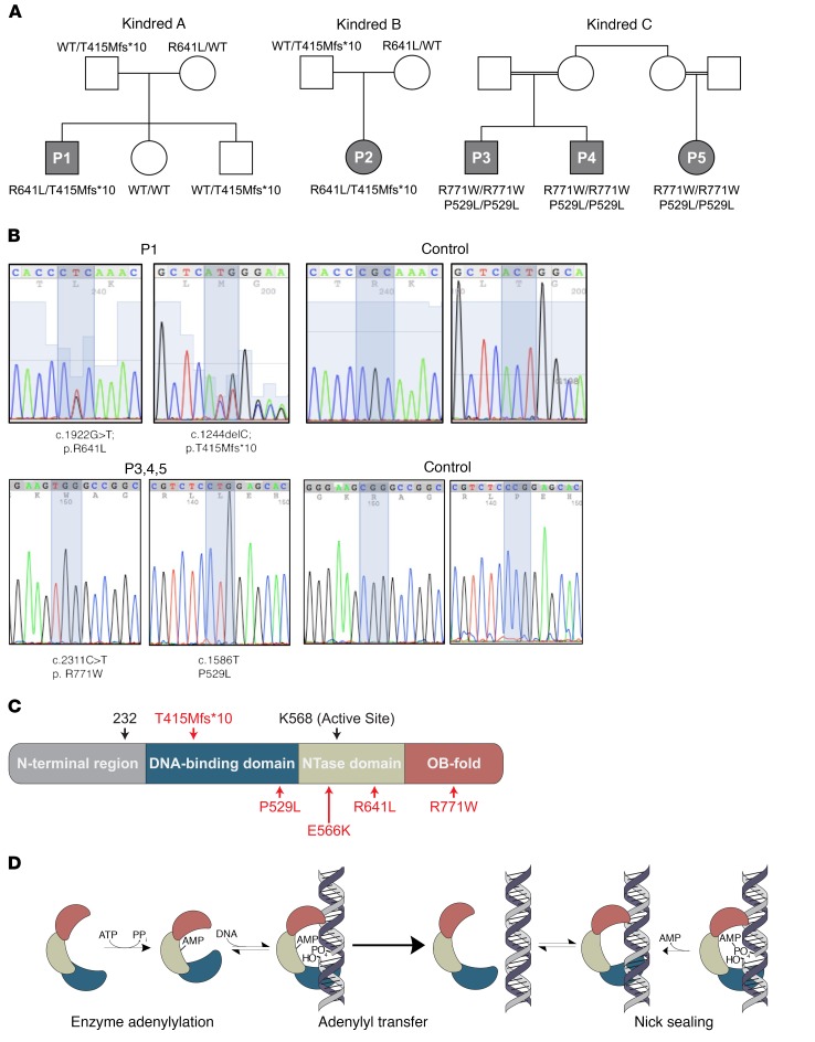
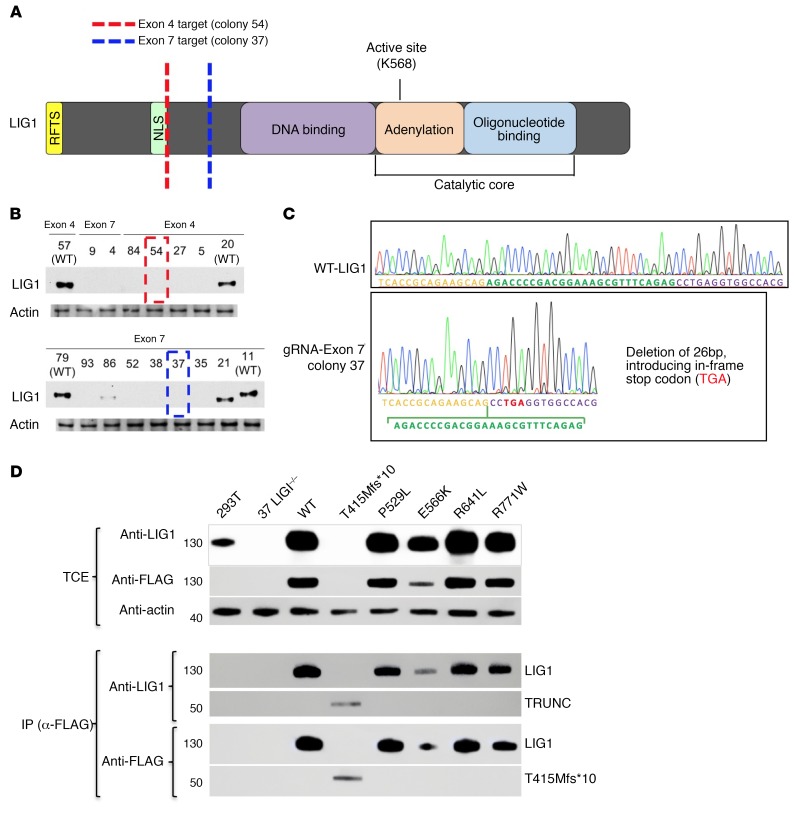
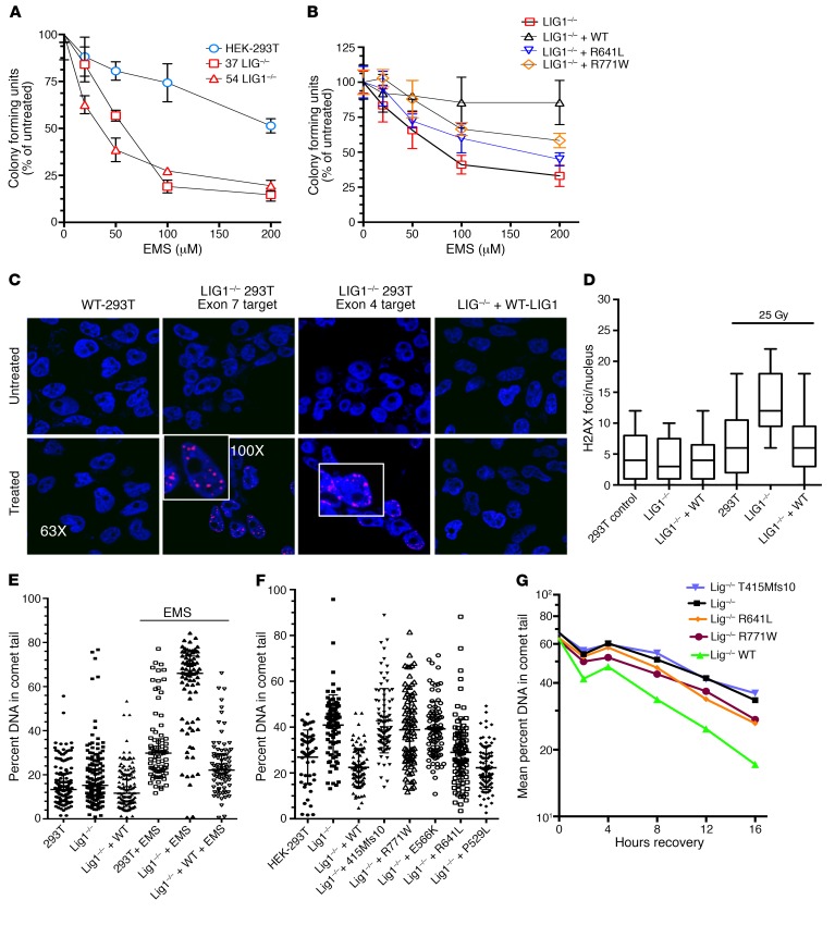
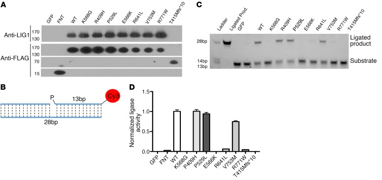
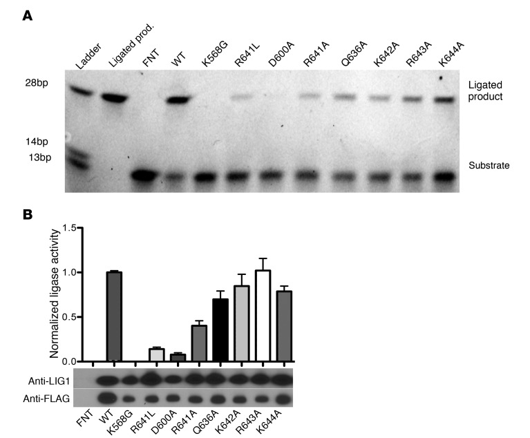
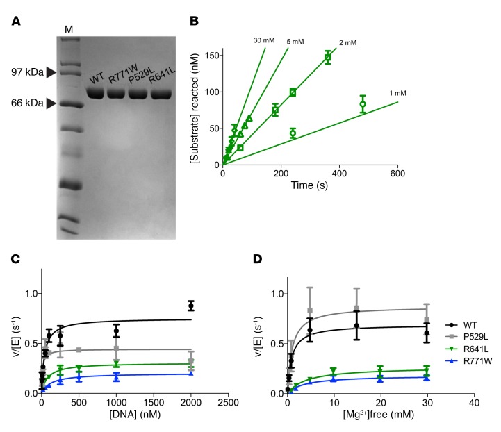
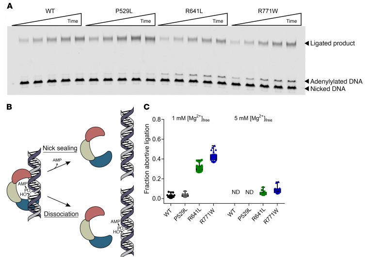
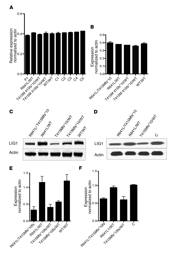
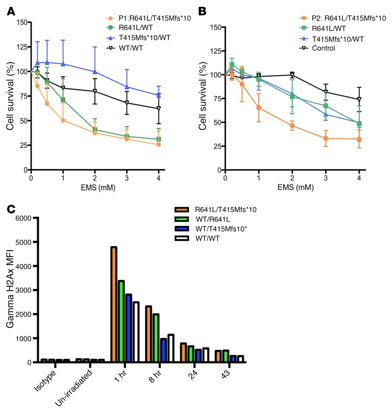
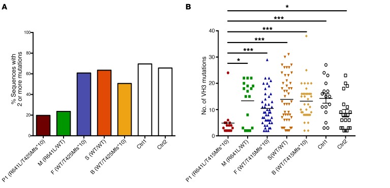
We report the molecular, cellular, and clinical features of 5 patients from 3 kindreds with biallelic mutations in the autosomal LIG1 gene encoding DNA ligase 1. The patients exhibited hypogammaglobulinemia, lymphopenia, increased proportions of circulating γδT cells, and erythrocyte macrocytosis. Clinical severity ranged from a mild antibody deficiency to a combined immunodeficiency requiring hematopoietic stem cell transplantation. Using engineered LIG1-deficient cell lines, we demonstrated chemical and radiation defects associated with the mutant alleles, which variably impaired the DNA repair pathway. We further showed that these LIG1 mutant alleles are amorphic or hypomorphic, and exhibited variably decreased enzymatic activities, which lead to premature release of unligated adenylated DNA. The variability of the LIG1 genotypes in the patients was consistent with that of their immunological and clinical phenotypes. These data suggest that different forms of autosomal recessive, partial DNA ligase 1 deficiency underlie an immunodeficiency of variable severity.

Keywords: B cells; Genetic diseases; Genetics; Immunology; T cell development.


Conflict of interest statement

Conflict of interest: The authors have declared that no conflict of interest exists.

Figures

Figure 1
Figure 1. LIG1 mutations and functions.
(A) Pedigrees for 3 kindreds with recessive mutations in LIG1. Patients 1 and 2 (P1 and P2) are from unrelated parents and both have the LIG1 mutations T415Mfs*10 and R641L. Patients 3 and 4 (P3 and P4) are brothers whose parents share grandmothers; patient 5 (P5) is a maternal cousin; these subjects have homozygous missense variants, P529L and R771W. The parents of P5 also share the same grandmother. (B) Sanger sequencing of P1 and a control, also P3 and a control. (Note that P2 has the same mutations as P1 and that P3 has the same genotype as P4 and P5.) (C) Schematic of LIG1 protein structure and approximate locations of all the identified mutations, in this and prior reports (16, 17). (D) Mechanism of LIG1-catalyzed DNA ligation. LIG1 consumes ATP and forms a covalent bond between AMP and K568. AMP is then transferred to the 5′-phosphate of a nicked DNA substrate. Attack by the 3′-hydroxyl seals the nick and releases AMP. All 3 chemical steps require Mg2+ as an essential cofactor.
Figure 2
Figure 2. Production of LIG1–/– and mutant cells.
(A) Schematic of LIG1 showing location of the gRNA targets. Targets were chosen in exon 4 (red) and exon 7 (blue), which are both upstream of the catalytic core. (B) Immunoblot using the rabbit polyclonal anti-LIG1 on cell lysates from CRISPR-Cas9–treated colonies. One colony from each exon (red and blue boxes) was chosen for experiments. (C) Sanger sequencing of exon 4 colony demonstrated the homozygous deletion of 32 bp and introduction of a premature stop codon; the exon 7 colony 37 showed homozygous deletion of 26 bp and also a premature stop codon. Plasmids containing WT LIG1 or the mutants found in LIG1 from patients (P529L, E566K, R771W, R641L, or T415Mfs*10) were transduced into colony 37 LIG1–/– HEK-293T cells using retroviral vectors. (D) Immunoblots of total cell lysates extracts (TCEs; performed 2 times) demonstrated absence of expression of LIG1 protein in the LIG1 knockout (clone 37) and restoration of LIG1 expression by transduced cells, as examined by rabbit polyclonal anti-LIG1. Immunoprecipitation with anti-FLAG demonstrated both FLAG and LIG1 polypeptide in these cells, validating the expression of truncated LIG1 in the T415Mfs*10 mutant. Each mutation was validated with Sanger sequencing (not shown).
Figure 3
Figure 3. Defective repair of LIG1–/– HEK-293T and mutant cells.
(A) LIG1–/– clones 37 and 54 were more sensitive to EMS, as compared with HEK-293T cells but similar to each other (2 experiments, 6 replicates; mean ± SD; P = 0.03, P = 0.04). (B) EMS sensitivity of clone 37 cells was rescued by complementation with WT LIG1, but only partially with R641L or R771W (3 experiments, 6 replicates; mean ± SD). (C) Responses of HEK-293T cells to γ radiation: WT HEK-293T cells, clone 37 LIG1–/–, and LIG1–/– + WT LIG1 were irradiated (25 Gy), incubated for 3 hours, and inspected for number of γH2AX foci per nuclei (3 experiments). (D) γH2AX foci/nucleus for HEK-293T cells, LIG1–/–, and LIG1–/– cells complemented with WT LIG1, before and after irradiation with 25 Gy. LIG1–/– cells demonstrated increased foci compared with HEK-293T cells (P = 0.0001), with substantial rescue with WT protein: LIG1–/– versus LIG1 –/– + WT, now P = 0.05 (3 experiments; 50 nuclei/condition counted per experiment; mean ± SD). (E) HEK-293T cells, LIG1–/– cells, and LIG–/– cells transfected with WT LIG1, incubated with media alone or 0.5 mM EMS, and examined by comet assay. Data are mean ± SD and percentage of cellular DNA in the comet tail for 79 to 86 cells/condition; performed 3 times. (F) Comparing DNA damage in 0.5 mM EMS-treated HEK-293T cells to LIG1–/–, T415Mfs*10, R771W, E566K cells, P < 0.0001, to R641L cells, P = 0.69. Adding the P529L variant was similar to adding WT LIG1 (differences, P = 0.9). (G) After 16 hours EMS, LIG1–/–, and mutant cells were washed, replated in fresh media, and tested for the differences in DNA damage at intervals. Mean percent shown for each. At 8 hours, differences emerged: HEK-293T versus LIG1–/–, P = 0.007; LIG1–/– versus T415Mfs*10, P = not significant; LIG1–/– versus R641L, P = 0.02; LIG1–/– versus R771W, P = 0.04 (50–88 colonies/condition).
Figure 4
Figure 4. Measurement of enzymatic activity of LIG1 mutants.
(A) Normalized expression of purified LIG1 protein from HEK-293T cells, containing patient or control mutations. Proteins were separated by SDS-PAGE and detected using rabbit polyclonal anti-LIG1 and anti-Flag antibodies as shown. (B) Schematic of 28 bp synthetic nicked oligonucleotide complex used for the enzymatic assay. (C) Representative ligation assay. Ligation forms a 28 bp ligated product. (D) Quantification of enzymatic activity normalized to WT LIG1 activity (%). (3 experiments; data are mean ± SD.) Controls: GFP and FNT= Flag, a nuclear localization signal, and thioredoxin.
Figure 5
Figure 5. Exploration of the DNA-binding loop containing Arg-641 by alanine scanning mutagenesis.
Residues adjacent to R641 in the DNA-binding loop were mutated to alanine (A) purified from HEK-293T cells, and then assessed for enzymatic activity. (A) Representative ligation assay. (B) Enzymatic activity is normalized to WT LIG1 activity (%), and the corresponding immunoblot is shown. Data are mean ± SD and are representative of 3 experiments using polyclonal anti-LIG1. Controls: WT LIG1; FNT= Flag, a nuclear localization signal, and thioredoxin.
Figure 6
Figure 6. Steady-state kinetic analysis of LIG1 mutants.
(A) Recombinant WT Δ232 LIG1 and mutants were purified to homogeneity and 1 μg protein analyzed by SDS-PAGE with Coomassie stain. M, marker. (B) Representative initial rates for ligation measured for R641L with varying amounts of Mg2+. The dependence on nicked DNA substrate (C) and Mg2+ cofactor (D) were determined for WT and mutant proteins (mean ± SD; n ≥ 3) and fit to the Michaelis-Menten equation (best fit parameters are summarized in Table 3).
Figure 7
Figure 7. R641L and R771W LIG1 mutants generate abortive ligation intermediates at physiological Mg2+ concentration.
(A) Representative denaturing polyacrylamide gel analyzing multiple-turnover ligation with 5 nM LIG1, 500 nM DNA, and 1 mM free Mg2+. (B) Schematic for abortive ligation by LIG1. After catalyzing adenylyl transfer, LIG1 can either catalyze the nick-sealing step or dissociate from the adenylylated DNA intermediate. (C) Fraction abortive ligation was calculated for WT and mutant enzymes at both 1 mM and 5 mM free Mg2+ (mean ± SD; n ≥ 3). ND, not detected. See Supplemental Figure 5 for the initial rates for formation of ligated product and adenylated DNA.
Figure 8
Figure 8. Protein and mRNA expression in B cells.
Reverse transcription qPCR was used to measure LIG1 mRNA levels in PBMCs (A) and EBV-B cells (B) from P1, family members, and 5 controls in 3 replicates. (C and D) EBV-B cell lysates were separated by SDS-PAGE and the expression of LIG1 was detected by chemiluminescence using rabbit monoclonal anti-LIG1 (recognizes aa 1–100) for P1 and family members (C) or P2 and family members with 1 control (D). (E) Quantification of LIG1 protein expression in blot C for P1, mother (R641L/WT), father (T415Mfs*10/WT), brother (T415Mfs*10/WT), and sister (WT/WT). (F) Quantification of LIG1 protein in blot D for P2, mother (R641L/WT) and father (T415Mfs*10/WT), as compared with a control (C) (immunoblotting, n = 3; data are mean ± SD).
Figure 9
Figure 9. Defective lymphocyte repair responses to EMS and radiation.
Lymphoblastoid B cells of subjects from kindred A (A) and kindred B (B) were cultured in media with increasing concentrations of ethyl methanesulfonate (EMS) to examine DNA sensitivity, determining cell survival (3 experiments; 6 replicates of cell cultures for each condition; mean ± SD). (C) The effects of radiation on peripheral blood T cells from P1 and family members were determined after exposure to 10 Gy radiation. After permeabilization, CD3+ T cell populations were examined by flow cytometer for γHAX foci, and the mean fluorescence intensity (MFI) was determined as described (43).
Figure 10
Figure 10. Effect of LIG1 deficiency on somatic hypermutation.
VH3 clones from 80 colonies per subject from P1 and family members (mother, father, sister, and brother) were amplified from cDNA from PBMC RNA, using primers specific for the VDJ VH3 region; individual clones were sequenced following TA-TOPO cloning. (A) The percentage of VH3 sequences in these clones for each member of the family that contained 2 or more mutations is shown, in comparison to similarly isolated clones of 2 healthy controls. (B) For clones that contained at least 2 mutations in the VH3 sequences, we then determined the number of mutations in each clone for each member of the family again, as compared with 2 healthy controls. *P = 0.05; ***P = 0.001.

Similar articles

Cited by

References

    1. Martin, MacNeill SA. ATP-dependent DNA ligases. Genome Biol. 2002;3(4):REVIEWS3005. - PMC - PubMed
    1. Grawunder U, Zimmer D, Fugmann S, Schwarz K, Lieber MR. DNA ligase IV is essential for V(D)J recombination and DNA double-strand break repair in human precursor lymphocytes. Mol Cell. 1998;2(4):477–484. doi: 10.1016/S1097-2765(00)80147-1. - DOI - PubMed
    1. van der Burg M, et al. A new type of radiosensitive T-B-NK+ severe combined immunodeficiency caused by a LIG4 mutation. J Clin Invest. 2006;116(1):137–145. doi: 10.1172/JCI26121. - DOI - PMC - PubMed
    1. Altmann T, Gennery AR. DNA ligase IV syndrome; a review. Orphanet J Rare Dis. 2016;11(1):137. - PMC - PubMed
    1. Simsek D, Jasin M. DNA ligase III: a spotty presence in eukaryotes, but an essential function where tested. Cell Cycle. 2011;10(21):3636–3644. doi: 10.4161/cc.10.21.18094. - DOI - PMC - PubMed

Publication types