. 2018 Dec 28:292:119-129.
doi: 10.1016/j.jconrel.2018.11.002. Epub 2018 Nov 2.

Design, formulation and evaluation of novel dissolving microarray patches containing a long-acting rilpivirine nanosuspension

Affiliations

Design, formulation and evaluation of novel dissolving microarray patches containing a long-acting rilpivirine nanosuspension

Maelíosa T C Mc Crudden et al. J Control Release. .

Abstract

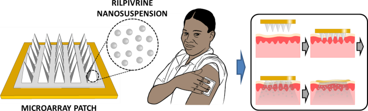
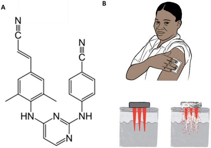
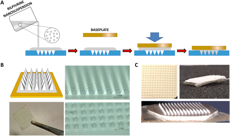
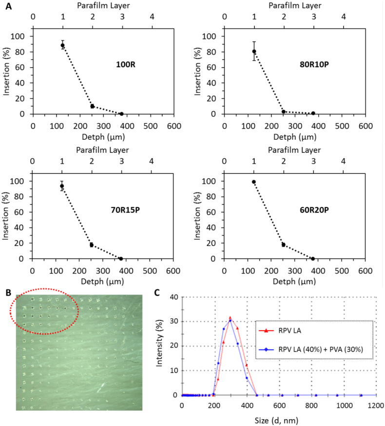
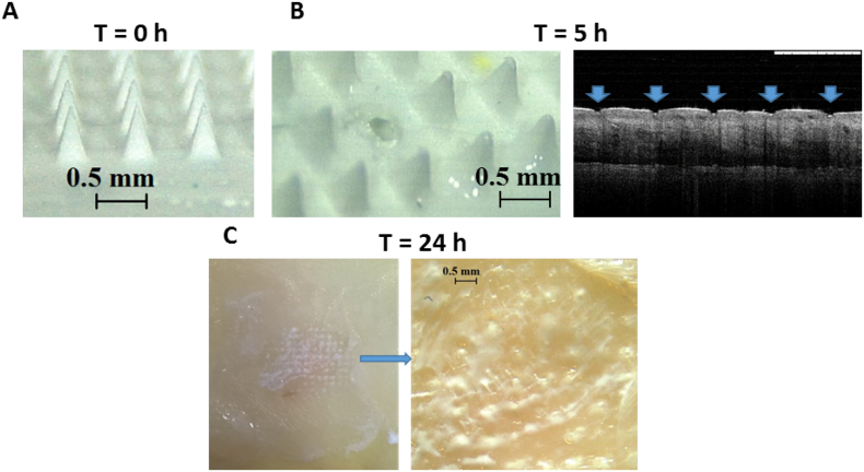
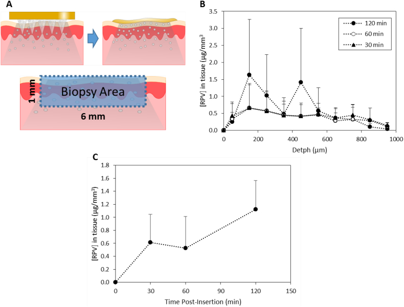
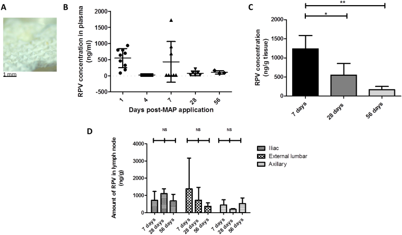
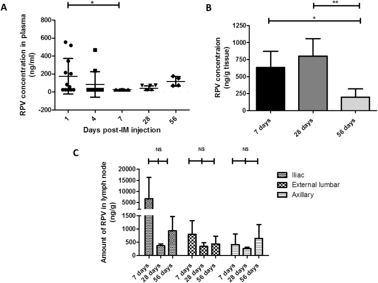
One means of combating the spread of human immunodeficiency virus (HIV) is through the delivery of long-acting, antiretroviral (ARV) drugs for prevention and treatment. The development of a discreet, self-administered and self-disabling delivery vehicle to deliver such ARV drugs could obviate compliance issues with daily oral regimens. Alternatives in development, such as long-acting intramuscular (IM) injections, require regular access to health care facilities and disposal facilities for sharps. Consequently, this proof of concept study was developed to evaluate the use of dissolving microarray patches (MAPs) containing a long-acting (LA) nanosuspension of the candidate ARV drug, rilpivirine (RPV). MAPs were mechanically strong and penetrated skin in vitro, delivering RPV intradermally. In in vivo studies, the mean plasma concentration of RPV in rats (431 ng/ml at the Day 7 time point) was approximately ten-fold greater than the trough concentration observed after a single-dose in previous clinical studies. These results are the first to indicate, by the determination of relative exposures between IM and MAP administration, that larger multi-array dissolving MAPs could potentially be used to effectively deliver human doses of RPV LA. Importantly, RPV was also detected in the lymph nodes, indicating the potential to deliver this ARV agent into one of the primary sites of HIV replication over extended durations. These MAPs could potentially improve patient acceptability and adherence to HIV prevention and treatment regimens and combat instances of needle-stick injury and the transmission of blood-borne diseases, which would have far-reaching benefits, particularly to those in the developing world.

Keywords: Antiretroviral; HIV; Microarray patch; Rilpivirine.


Figures

Unlabelled Image
Graphical abstract
Fig. 1
Fig. 1
The chemical structure of RPV (A). Schematic representation of the mode of RPV LA MAP application and subsequent in-skin dissolution of the RPV LA MAP (B).
Fig. 2
Fig. 2
Schematic representation of the RPV LA MAP manufacturing process (A); micrographs of exemplar RPV LA MAPs generated (B) and digital images of RPV LA MAPs (C).
Fig. 3
Fig. 3
Insertion profile of RPV LA MAPs containing different amounts of PVA and RPV LA into a Parafilm® multi-layered skin simulant, following the application of a pressure of 10 N/MAP, (means ± SD, n = 3) (A). Digital image of the first layer of the skin simulant after the insertion of MAP formulated containing no PVA (B). Size of RPV nanoparticles in nanosuspension (RPV Nanosusp) and RPV nanoparticles following MAP formulation with PVA, drying and then dissolution of the MAP in water (RPV Nanosusp (40%) + PVA (30%)) (C).
Fig. 4
Fig. 4
Representative digital micrographs illustrative of the dissolution of RPV LA MAP at specific time points (0, 5 and 24 h) in excised neonatal porcine skin. Digital micrograph of a RPV LA MAP prior to insertion into skin (A). Digital micrograph of a RPV LA MAP upon removal from the skin after an insertion time of 5 h and OCT image of the resulting microconduits in the porcine skin following MAP removal (B). Digital image and micrograph of porcine skin upon removal of RPV LA MAP post-24 h insertion with RPV LA visible in the microconduits which were created in the skin (C).
Fig. 5
Fig. 5
Insertion of RPV LA MAP into excised porcine skin, followed by deposition studies. RPV LA MAPs were inserted, for three different durations (30, 60 or 120 min), into excised full-thickness neonatal porcine skin, and following this, the deposition of drug into a biopsied piece of skin was determined. Schematic representation of the experimental approach, indicating diffusion of drug into the excised skin (A). RPV concentrations (μg/mm3) in excised neonatal porcine skin were determined, at the relevant time points, and as a function of the depth of the tissue sections. This experiment was carried out under temperature control at 37 °C, (means ± SD, n = 3) (B). The total amount of RPV (μg/mm3) in the biopsied tissue was expressed as a function of the application time (means ± SD, n = 3) (C).
Fig. 6
Fig. 6
RPV quantification in in vivo samples from animals treated with 4 × RPV LA MAPs (4 × 2 mg RPV LA) (MAP cohort, three rats for each time point). An exemplar MAP post-removal from a rat, indicating the extent of MAP dissolution over the course of the 24 h application period (A). Plasma levels of RPV at 1, 4, 7, 28 and 56 days post-MAP application, (means ± SD, n ≥ 3 at each time point, in accordance with the experimental regime employed) (B). No plasma at the 4 day time point had RPV levels above the LOQ of the system and so for analysis purposes, samples were treated as 25 ng/ml. Determination of the amount of RPV in excised vaginal tissue, expressed as ng of drug per g of vaginal tissue (ng/g), (means ± SD, n = 3; * P < 0.05; NS = not significant) (C). Determination of the amount of RPV in excised lymph nodes, expressed as ng of drug per g of lymph node tissue (ng/g) in animals treated for 7, 28 or 56 days, (means ± SD, n = 3 in all cases; NS = not significant) (D). No axillary nodes at the 28-day time point or external lumbar nodes at the 56-day time point had RPV levels above the LOQ of the system. For analysis purposes, these samples were all treated as 25 ng/ml and then adjusted for tissue mass.
Fig. 7
Fig. 7
RPV quantification in in vivo samples from animals treated with RPV LA via IM injection (1.8 mg) (IM cohort, four rats for each time point). Plasma levels of RPV at 1, 4, 7, 28 and 56 days post-IM injection of RPV, (means ± SD, n ≥ 4 at each time point, in accordance with the experimental regime employed) (A). No plasma at the 7-day time point had RPV levels above the LOQ of the system and so for analysis purposes, samples were treated as 25 ng/ml. Determination of the amount of RPV in excised vaginal tissue, expressed as ng of drug per g of vaginal tissue (ng/g), (means ± SD, n = 4; * P < 0.05; NS = not significant) (B). Determination of the amount of RPV in excised lymph nodes, expressed as ng of drug per g of lymph node tissue (ng/g) in animals treated for 7, 28 or 56 days, (means ± SD, n = 4 in all cases; NS = not significant) (C). No lymph nodes at the 28-day time point had RPV levels above the LOQ of the system. For analysis purposes, these samples were all treated as 25 ng/ml and then adjusted for tissue mass.

Similar articles

Cited by

References

    1. Public Health England HIV diagnoses, late diagnoses and numbers accessing treatment and care. Version. 2016;1(0):4–8.
    1. Mosam A., Dlova N.C. HIV/AIDS in sub-Saharan Africa. Dermatol. Clin. 2006;24(4):421–429. - PubMed
    1. Sawers L., Isaac A. Partnership duration, concurrency, and HIV in sub-Saharan Africa. Afr. J. AIDS Res. 2017;17:1–10. - PubMed
    1. Nyaku A.N., Kelly S.G., Taiwo B.O. Long-acting antiretrovirals: where are we now? Curr. HIV/AIDS Rep. 2017;14(2):63–71. - PubMed
    1. Azijn H. TMC278, a next generation nonnucleoside reverse transcriptase inhibitor (NNRTI), active against wild type and NNRTI-resistant HIV-1. Antimicrob. Agents Chemother. 2010;54(2):718–727. - PMC - PubMed

Publication types