. 2019 Jan 2:526:173-179.
doi: 10.1016/j.virol.2018.10.021. Epub 2018 Nov 2.

Vascular permeability in the brain is a late pathogenic event during Rift Valley fever virus encephalitis in rats

Affiliations

Vascular permeability in the brain is a late pathogenic event during Rift Valley fever virus encephalitis in rats

Aaron W Walters et al. Virology. .

Abstract

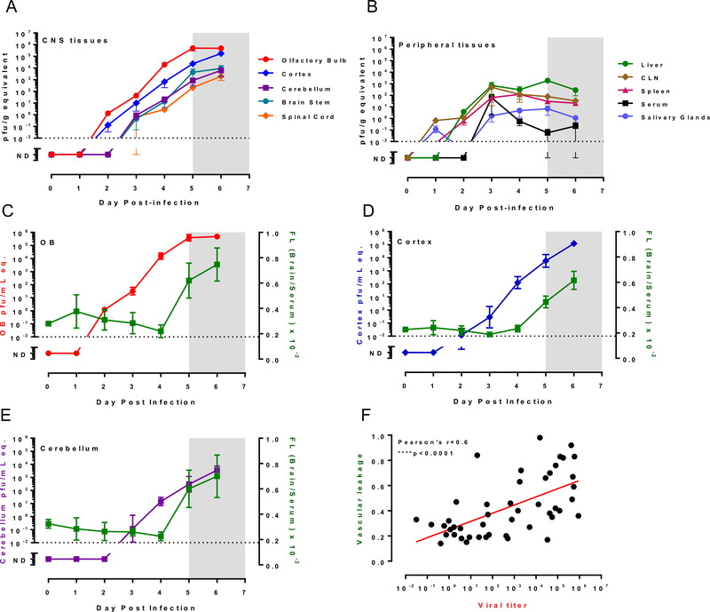
Rift Valley fever virus (RVFV) is a zoonotic disease of livestock that causes several clinical outcomes in people including febrile disease, hemorrhagic fever, and/or encephalitis. After aerosol infection with RVFV, Lewis rats develop lethal encephalitic disease, and we use this as a model for studying disease mechanisms of RVFV infection in the brain. Permeability of the brain vasculature in relation to virus invasion and replication is not known. Here, we found that vascular permeability in the brain occurred late in the course of infection and corresponded temporally to expression of matrix metalloproteinase-9 (MMP-9). Virus replication was ongoing within the central nervous system for several days prior to detectable vascular leakage. Based on this study, vascular permeability was not required for entry of RVFV into the brain of rats. Prevention of vascular leakage late in infection may be an important component for prevention of lethal neurological disease in the rat model.

Keywords: Aerosol infection; Blood brain barrier; Rift valley fever; Viral encephalitis.


Figures

Figure 1.
Figure 1.. Virus replication and vascular breakdown during RVF encephalitis.
Lewis rats were infected with 1×10³ pfu of RVFV by aerosol and then serially euthanized (n=3) over a time course of six days. (A-B) Virus levels in tissues and serum were measured by q-RT-PCR and compared to virus stock of known titer to generate pfu/g or pfu/ml equivalent (y-axes). (A) CNS tissues; (B) peripheral tissues. CLN = cervical lymph nodes. (C-E) Fluorescein-salt penetration of brain regions was normalized to serum (right y-axis; green). Corresponding viral q-RT-PCR results for each sample are normalized to plaque forming units and are represented as pfu/g equivalents (left y-axis). The limit of detection of the PCR assay is indicted by the horizontal dotted line. ND = below the limit of detection. The gray shaded area between 5–7 dpi represents the clinical window when rats display clinical signs of disease. (F) Vascular leakage (normalized fluorescent intensity) of all brain samples were graphed against the viral RNA from each corresponding sample. Red line indicates linear regression of viral titer and vascular leakage. Pearson’s correlation was performed with r and p values indicated on graph.
Figure 2.
Figure 2.. In vivo imaging of vascular integrity in RVFV-infected rats.
Lewis rats were exposed to 2×10³ pfu of RVFV. At 4 dpi, a pair of rats was injected with Superhance 680 then imaged for fluorescent intensity at 5 min, 3 hr, 24 hr, and 48 hr post-agent injection. This figure represents 1 pair of rats: uninfected control (-CTL; left) and an infected rat (RVF+; right) featuring both the dorsal and ventral views. After euthanasia on day 6, the brains were extracted and imaged ex vivo (bottom row). Radiant efficiency was normalized across samples and the scale is included.
Figure 3.
Figure 3.. Vascular integrity is compromised in lethally infected rats.
(A) Four additional pairs of rats were imaged as described in Fig. 2. Each pair consists of an uninfected control (left) and infected (right) rat imaged 24 hr after Superhance injection. Pairs 3 and 4 show the corresponding extracted brain. (B) Quantification of vascular leakage as determined by IVIS. Statistical significance indicated by asterisks. N=5 for the whole-head images from live rats; n=3 for the extracted brains.
Figure 4.
Figure 4.. Levels of MMP-9 correlate with viral titer.
Lewis rats were infected with 1×10⁴ pfu of RVFV by aerosol and then serially euthanized (n=3) over a time course of six days. (A) MMP-9 levels (right y-axis) were measured in serum (gray) and brain cortex (blue) samples. Corresponding viral RNA in the brain is shown in pfu/g equivalents (red; left y-axis). The limit of detection of the PCR assay is indicted by the horizontal dotted line. The gray shaded area between 5–7 dpi represents the clinical window when rats display clinical signs of disease. (B) MMP-9 levels in brain samples were graphed against the viral RNA from each corresponding sample. Lines indicate linear regression of viral titer and MMP-9 levels in serum (gray) and cortex (blue). Pearson’s correlation was performed for cortex samples with r and p values indicated on graph.

References

    1. Al-Hazmi M, Ayoola EA, Abdurahman M, Banzal S, Ashraf J, El-Bushra A, Hazmi A, Abdullah M, Abbo H, Elamin A, Al-Sammani el T, Gadour M, Menon C, Hamza M, Rahim I, Hafez M, Jambavalikar M, Arishi H, Aqeel A, 2003. Epidemic Rift Valley fever in Saudi Arabia: a clinical study of severe illness in humans. Clin Infect Dis 36, 245–252. - PubMed
    1. Anyangu AS, Gould LH, Sharif SK, Nguku PM, Omolo JO, Mutonga D, Rao CY, Lederman ER, Schnabel D, Paweska JT, Katz M, Hightower A, Njenga MK, Feikin DR, Breiman RF, 2010. Risk factors for severe Rift Valley fever infection in Kenya, 2007. Am J Trop Med Hyg 83, 14–21. - PMC - PubMed
    1. Bales JM, Powell DS, Bethel LM, Reed DS, Hartman AL, 2012. Choice of inbred rat strain impacts lethality and disease course after respiratory infection with Rift Valley Fever Virus. Frontiers in cellular and infection microbiology 2, 105. - PMC - PubMed
    1. Bird BH, Reynes JM, Nichol ST, 2012. Rift Valley Fever, in: Magill AJ, Strickland GT, Maguire JH, Ryan ET, Solomon T (Eds.), Hunter’s Tropical Medicine and Emerging Infectious Disease, 9th ed. Elsevier Health Sciences, pp. 340–343.
    1. Boushab BM, Fall-Malick FZ, Baba SEWO, Salem MLO, Belizaire MRD, Ledib H, Ahmed MMOB, Basco LK, Ba H, 2016. Severe human illness caused by rift valley fever virus in Mauritania, 2015. Open Forum Infectious Diseases 3. - PMC - PubMed

Publication types

Substances