

Conus Medullaris Levels on Ultrasonography in Term Newborns : Normal Levels and Dermatological Findings
- PMID: 30396246
- PMCID: PMC6280053
- DOI: 10.3340/jkns.2016.1212.001
Conus Medullaris Levels on Ultrasonography in Term Newborns : Normal Levels and Dermatological Findings
Abstract
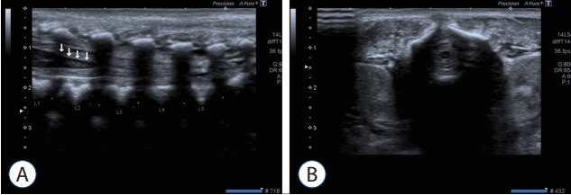
Objective: Ultrasonography (US) is the most non-invasive, safe, and, especially in the period of infancy, best method for visualizing and examining the spinal cord. Furthermore, US is the primary work-up for development of the spinal canal, and for follow-up on issues relating to subcutaneous tissues, bone development, and the spinal cord. Conus medullaris terminates at the second lumbar vertebra, according to a consensus in the literature.
Methods: Healthy children under the age of 6 months who were admitted to the radiology clinic for routine USG follow-ups between the dates of March 2012 to December 2014 were included in this study.
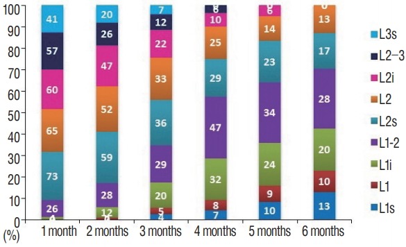
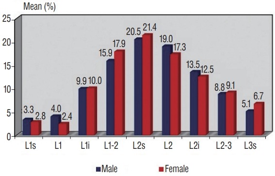
Results: Our study includes data from 1125 lumbosacral ultrasounds. The terminal point of the conus level of the attended infants, superior, middle part, inferior of the vertebrae L1, L2, and L3. Furthermore, the termination of the discal distance ratio did not differ significantly between genders.
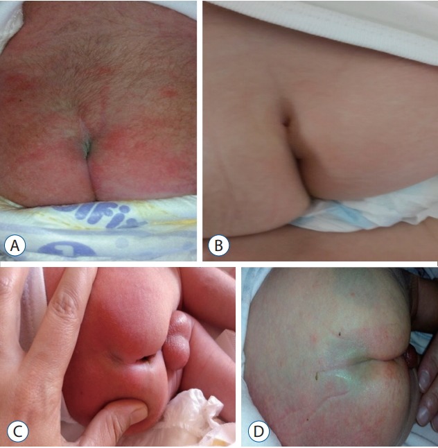
Conclusion: Therefore, according to our results, gender is not an influencing factor in the termination of the spinal cord. Based on the study we performed, as well as the previous literature, in infants without a recognized spinal pathology, the spinal cord is detected below the vertebra L3.
Keywords: Conus medullaris; Term birth; Ultrasonography.
Conflict of interest statement
No potential conflict of interest relevant to this article was reported.
Figures
References
-
- Arthurs OJ, Thayyil S, Wade A, Chong WK, Sebire NJ, Taylor AM, Magnetic Resonance Imaging Autopsy Study Collaborative Group Normal ascent of the conus medullaris: a post-mortem foetal MRI study. J Matern Fetal Neonatal Med. 2013;26:697–702. - PubMed
-
- Değirmenci S, Güven F, Celayir A, Kılıç BD, Say A. Lumbosakral orta hat cilt lezyonlu yenidoğanlarda spinal kord anomalileri Orijinal Araştırma. Türk Pediatri Arşivi. 2003;38:103–106.
-
- Ferahbas A, Utas S, Akcakus M, Gunes T, Mistik S. Prevalence of cutaneous findings in hospitalized neonates: a prospective observational study. Pediatr Dermatol. 2009;26:139–142. - PubMed
-
- Gokdemir G, Erdogan HK, Köşlü A, Baksu B. Cutaneous lesions in Turkish neonates born in a teaching hospital. Indian J Dermatol Venereol Leprol. 2009;75:638. - PubMed
-
- Henriques JG, Pianetti G, Henriques KS, Costa P, Gusmão S. Minor skin lesions as markers of occult spinal dysraphisms--prospective study. Surg Neurol. 2005;63:8–12. - PubMed
LinkOut - more resources
Full Text Sources
