Review
. 2019 Apr 1;11(4):a033894.
doi: 10.1101/cshperspect.a033894.

Early Events in the Endoplasmic Reticulum Unfolded Protein Response

Affiliations
Review

Early Events in the Endoplasmic Reticulum Unfolded Protein Response

Steffen Preissler et al. Cold Spring Harb Perspect Biol. .

Erratum in

Abstract

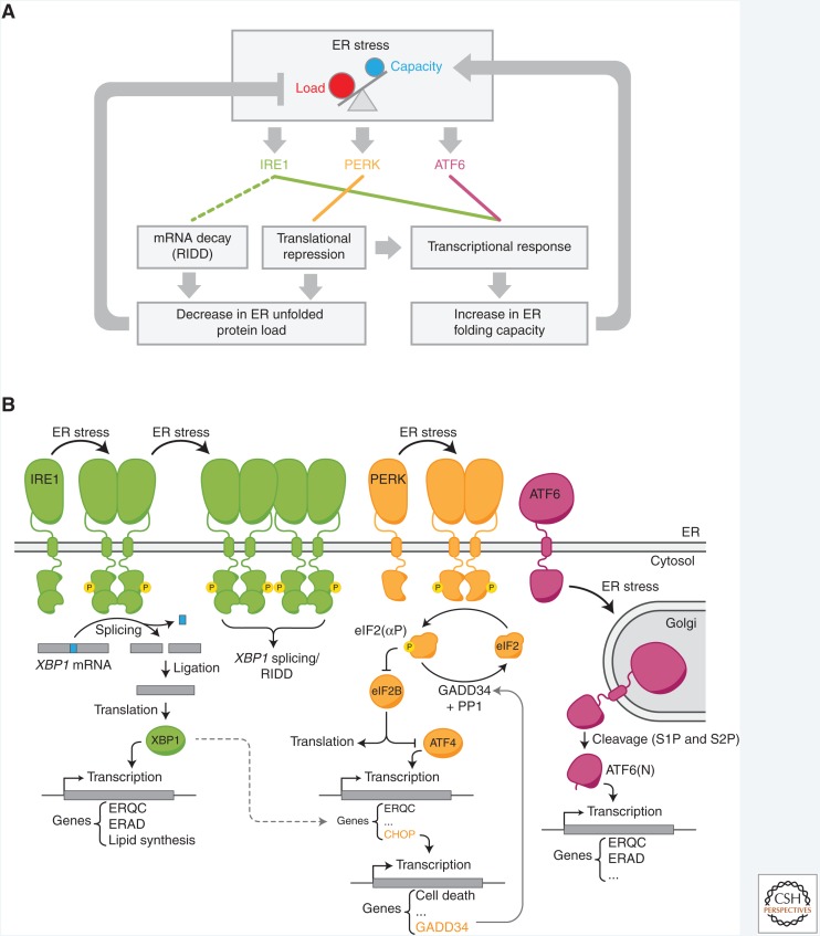
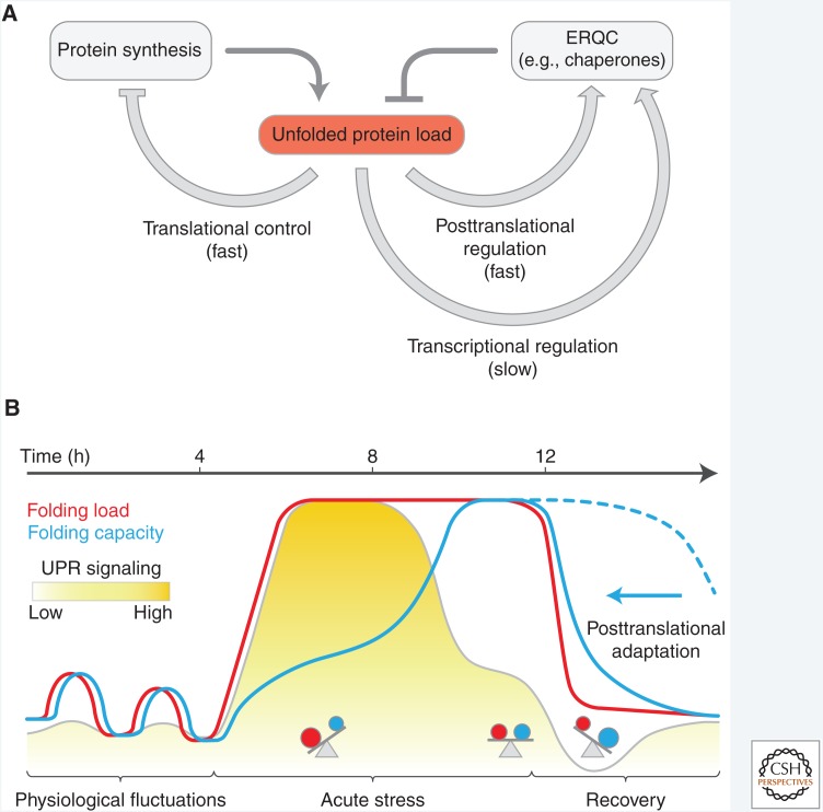
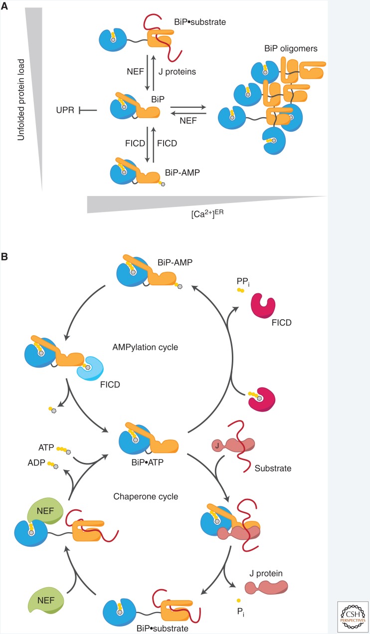
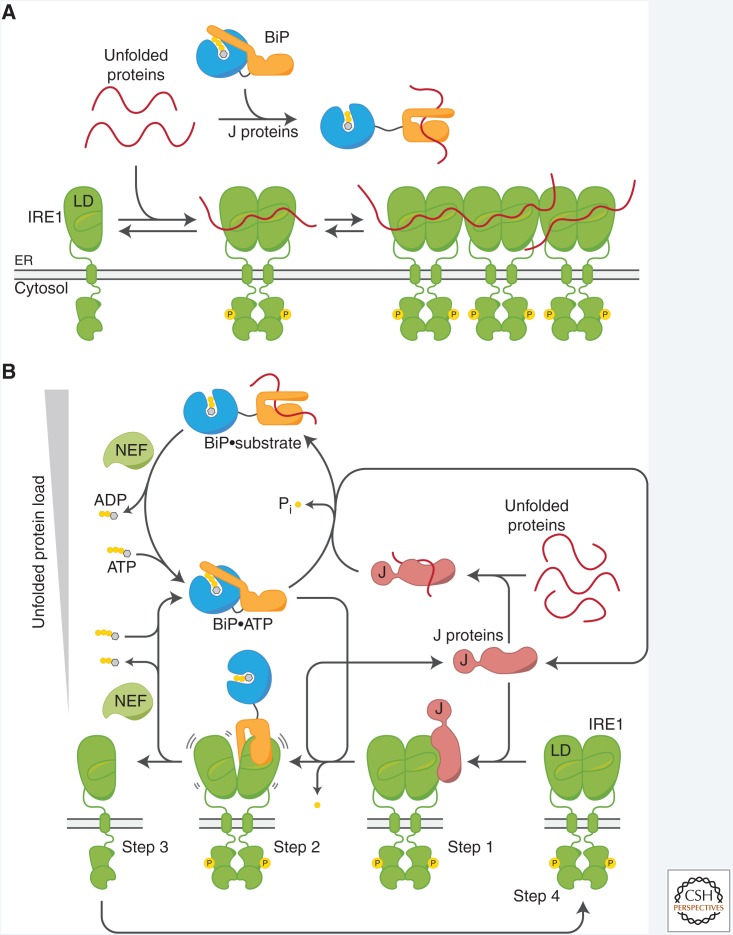
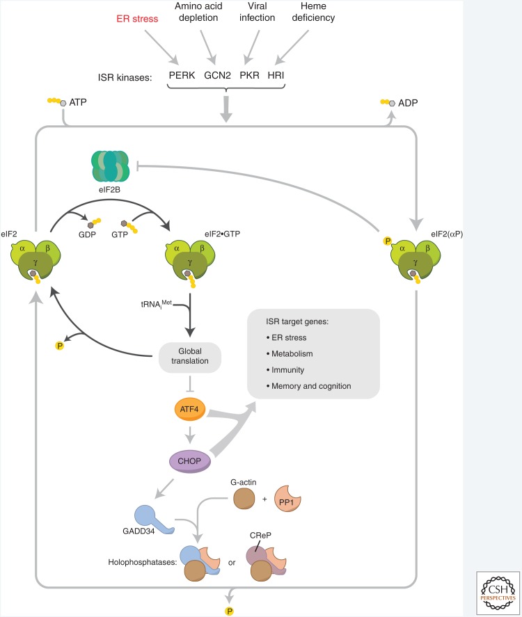
The physiological consequences of the unfolded protein response (UPR) are mediated by changes in gene expression. Underlying them are rapid processes involving preexisting components. We review recent insights gained into the regulation of the endoplasmic reticulum (ER) Hsp70 chaperone BiP, whose incorporation into inactive oligomers and reversible AMPylation and de-AMPylation present a first line of response to fluctuating levels of unfolded proteins. BiP activity is tied to the regulation of the UPR transducers by a recently discovered cycle of ER-localized, J protein-mediated formation of a repressive IRE1-BiP complex, whose working we contrast to an alternative model for UPR regulation that relies on direct recognition of unfolded proteins. We conclude with a discussion of mechanisms that repress messenger RNA (mRNA) translation to limit the flux of newly synthesized proteins into the ER, a rapid adaptation that does not rely on new macromolecule biosynthesis.


Figures

Figure 1.
Figure 1.
Overview of the mammalian unfolded protein response (UPR). (A) The central elements of the UPR signaling network. Each of the three signal transducers: IRE1, protein kinase R-like endoplasmic reticulum (ER) kinase (PERK), and activating transcription factor 6 (ATF6) constitutes a separate branch of the UPR with a specific output to counteract proteostatic perturbations in the ER. (B) Basic components and downstream events of the UPR signaling pathways. ER stress induces dimerization/oligomerization of IRE1 and its trans-autophosphorylation, which leads to nonconventional XBP1 messenger RNA (mRNA) splicing and production of the functional XBP1 transcription factor to induce expression of genes involved in ER protein quality control (ERQC) and ER-associated degradation (ERAD) of misfolded proteins. Activated, oligomeric IRE1 may also degrade ER-targeted mRNAs (via regulated Ire1-dependent decay [RIDD]). PERK-mediated phosphorylation of the α subunit of the eukaryotic initiation factor 2 (eIF2α) causes general translation attenuation, to relieve the load of newly synthesized proteins, and preferred synthesis of ATF4 (see Fig. 5). Production of the downstream transcription factor C/EBP homologous protein (CHOP) feeds back negatively on the PERK pathway (by enhancing expression of the phosphorylated eIF2 [eIF2(αP)]-specific regulatory phosphatase subunit growth arrest and DNA damage 34 [GADD34]) and induction of proapoptotic genes to favor cell death at later stages of unrectified stress. ER stress also induces ATF6 translocation to the Golgi where it is processed by S1P and S2P proteases to liberate the ATF6(N) transcription factor element. At the transcriptional level, all branches primarily facilitate expansion of the ER folding capacity and crosstalk exists between them (e.g., dotted line).
Figure 2.
Figure 2.
Latency of the negative feedback-regulated transcriptional unfolded protein response (UPR). (A) Adaptation of the UPR by negative feedback regulation via endoplasmic reticulum (ER) protein quality control (ERQC) components. Apart from stress-denatured proteins, newly synthesized secretory proteins constitute the majority of unfolded species in the ER and their accumulation activates the UPR transducers. The response entails rapid attenuation of translation to alleviate the unfolded protein load (see Fig. 5), rapid posttranslational regulation of the pool of active chaperones in the ER (see Fig. 3), and the slower induction of ERQC genes to expand the folding capacity and mediate negative feedback regulation of the UPR (see Fig. 4). (B) Theoretical, time-resolved response profiles to illustrate the inherent latency of the UPR. Recurring physiological short-term fluctuations in the unfolded protein load do not cause a significant transcriptional response, suggesting that they are buffered by rapid, nontranscriptional adjustments of the folding capacity. During acute stress the fast rise of the folding load induces UPR signaling but expansion of the folding capacity is delayed as a result of the inherent latency of transcriptional reprogramming. Negative feedback inhibition attenuates the transcriptional response as the folding capacity builds up to establish homeostasis at a new level. During the recovery from ER stress the folding load drops rapidly. Were the adjustment in ERQC capacity to rely exclusively on the decay of the ERQC messenger RNAs (mRNAs) and encoded proteins, ERQC capacity would likely exceed the unfolded protein load in this recovery phase (dotted blue line). However, posttranslational mechanisms that inactivate chaperones rapidly adjust the effective folding capacity and prevent over-chaperoning (arrow).
Figure 3.
Figure 3.
Posttranslational regulation of BiP. (A) Dynamic partitioning of BiP among different pools. Unfolded substrate polypeptides directly compete with oligomerization for a limited pool of free, ATP-bound BiP. Free BiP also interacts with unfolded protein response (UPR) signal transducers and contributes to repression of the UPR (see Fig. 4). J proteins promote both high-affinity substrate interactions and competing BiP oligomerization (with yet-to-be-characterized relative kinetics), whereas nucleotide exchange factors (NEFs) induce substrate release and oligomer disassembly. When the folding load declines, ATP-bound BiP becomes reversibly modified and inactivated by FICD-mediated AMPylation. Calcium depletion from the endoplasmic reticulum (ER) strongly induces BiP oligomerization by an unknown mechanism. (B) Chaperone and AMPylation cycles of BiP. ATP-bound BiP can be recruited to its substrates by J proteins, which stimulate ATP hydrolysis by BiP to achieve high-affinity interactions with its substrates. NEFs promote ADP release from BiP to enable ATP rebinding and dissociation of substrates. When the folding load is low, ATP-bound BiP becomes AMPylated by FICD. This locks BiP in a low-affinity state for substrates and renders the chaperone insensitive to stimulation by J proteins, thereby causing its functional inactivation. The same enzyme, FICD, demodifies BiP when the unfolded protein load increases to rapidly recruit preexisting inactive BiP into the pool of active chaperones. The different functional states of FICD, default de-AMPylation and yet-to-be-defined AMPylation, are indicated in light blue and magenta, respectively.
Figure 4.
Figure 4.
Role of BiP in regulating IRE1. (A) Direct binding model. Accumulating unfolded proteins bind directly to a groove on the luminal domain (LD) of IRE1 to promote its dimerization/oligomerization and activation. In this model, sequestration of unfolded proteins by chaperones such as BiP counteracts their binding to IRE1, repressing the unfolded protein response (UPR) and establishing a measure for the balance between unfolded proteins and available chaperones. (B) Chaperone inhibition model. Step 1: Endoplasmic reticulum (ER)-localized J proteins (exemplified by ERdj4) associate with the LD of (dimeric) IRE1. Step 2: The J protein recruits BiP and stimulates its ATPase activity to promote a canonical substrate interaction between BiP and IRE1. Step 3: BiP binding triggers J protein release and disrupts IRE1 dimers, causing IRE1 inactivation. BiP dissociates from IRE1 upon nucleotide exchange. Step 4: Chaperone-free IRE1 has an intrinsic propensity to dimerize and become active. J protein–mediated loading of BiP onto IRE1 competes with loading of BiP onto unfolded substrates. The availability of BiP (and J proteins) establishes a dynamic pool of monomeric, inactive IRE1.
Figure 5.
Figure 5.
The integrated stress response (ISR). Translational control over client protein load is imposed by the endoplasmic reticulum (ER) stress-mediated PERK activation and phosphorylation of eIF2 on its α subunit. This figure highlights the inhibition the guanine nucleotide exchange factor (GEF), eIF2B, by phosphorylated eIF2 and the effects on messenger RNA (mRNA) translation of the resultant decline in availability of eIF2•GTP•tRNAiMet ternary complexes. Also cartooned are the phosphatase complexes that dephosphorylate eIF2 to terminate signaling in the ISR. Targeting this phosphatase complexes for pharmacological inhibition has emerged as a potentially useful way to combat certain diseases of protein misfolding, by enhancing ISR activity.

Similar articles

Cited by

References

    1. Abastado JP, Miller PF, Jackson BM, Hinnebusch AG. 1991. Suppression of ribosomal reinitiation at upstream open reading frames in amino acid-starved cells forms the basis for GCN4 translational control. Mol Cell Biol 11: 486–496. - PMC - PubMed
    1. Abravaya K, Myers MP, Murphy SP, Morimoto RI. 1992. The human heat shock protein hsp70 interacts with HSF, the transcription factor that regulates heat shock gene expression. Genes Dev 6: 1153–1164. - PubMed
    1. Adamson B, Norman TM, Jost M, Cho MY, Nunez JK, Chen Y, Villalta JE, Gilbert LA, Horlbeck MA, Hein MY, et al. 2016. A multiplexed single-cell CRISPR screening platform enables systematic dissection of the unfolded protein response. Cell 167: 1867–1882, e1821. - PMC - PubMed
    1. Amin-Wetzel N, Saunders RA, Kamphuis MJ, Rato C, Preissler S, Harding HP, Ron D. 2017. A J-protein co-chaperone recruits BiP to monomerize IRE1 and repress the unfolded protein response. Cell 171: 1625–1637. - PMC - PubMed
    1. Axten JM, Medina JR, Feng Y, Shu A, Romeril SP, Grant SW, Li WH, Heerding DA, Minthorn E, Mencken T, et al. 2012. Discovery of 7-Methyl-5-(1-{[3-(trifluoromethyl)phenyl]acetyl}-2,3-dihydro-1H-indol-5-yl)-7H-pyrrolo[2,3-d]pyrimidin-4-amine (GSK2606414), a potent and selective first-in-class inhibitor of protein kinase R (PKR)-like endoplasmic reticulum kinase (PERK). J Med Chem 55: 7193–7207. - PubMed

Publication types

MeSH terms

Substances

LinkOut - more resources