. 2019 Jan 24;87(2):e00628-18.
doi: 10.1128/IAI.00628-18. Print 2019 Feb.

Rickettsia rickettsii Whole-Cell Antigens Offer Protection against Rocky Mountain Spotted Fever in the Canine Host

Affiliations

Rickettsia rickettsii Whole-Cell Antigens Offer Protection against Rocky Mountain Spotted Fever in the Canine Host

Andy Alhassan et al. Infect Immun. .

Abstract

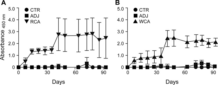
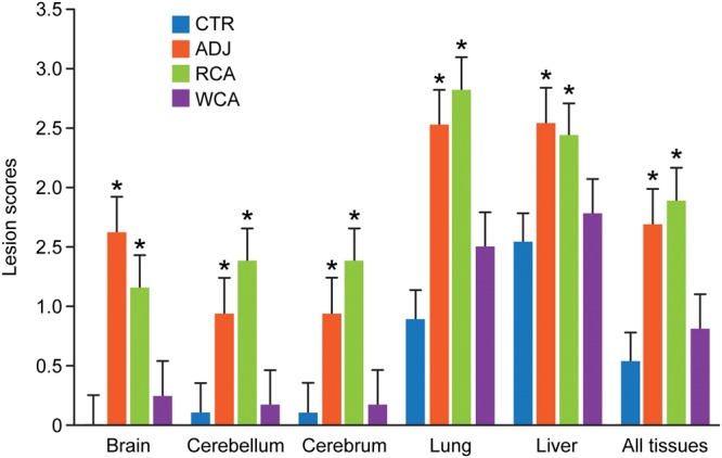
Rocky Mountain spotted fever (RMSF) is a potentially fatal tick-borne disease in people and dogs. RMSF is reported in the United States and several countries in North, Central, and South America. The causative agent of this disease, Rickettsia rickettsii, is transmitted by several species of ticks, including Dermacentor andersoni, Rhipicephalus sanguineus, and Amblyomma americanum RMSF clinical signs generally include fever, headache, nausea, vomiting, muscle pain, lack of appetite, and rash. If untreated, it can quickly progress into a life-threatening illness in people and dogs, with high fatality rates ranging from 30 to 80%. While RMSF has been known for over a century, recent epidemiological data suggest that the numbers of documented cases and the fatality rates remain high in people, particularly during the last two decades in parts of North America. Currently, there are no vaccines available to prevent RMSF in either dogs or people. In this study, we investigated the efficacies of two experimental vaccines, a subunit vaccine containing two recombinant outer membrane proteins as recombinant antigens (RCA) and a whole-cell inactivated antigen vaccine (WCA), in conferring protection against virulent R. rickettsii infection challenge in a newly established canine model for RMSF. Dogs vaccinated with WCA were protected from RMSF, whereas those receiving RCA developed disease similar to that of nonvaccinated R. rickettsii-infected dogs. WCA also reduced the pathogen loads to nearly undetected levels in the blood, lungs, liver, spleen, and brain and induced bacterial antigen-specific immune responses. This study provides the first evidence of the protective ability of WCA against RMSF in dogs.

Keywords: RMSF; Rickettsia; Rocky Mountain spotted fever; tick-borne pathogens; vaccines; vector-borne diseases.


Figures

FIG 1
FIG 1
R. rickettsii-specific IgG response following vaccination and infection challenge. Antigen-specific IgG was measured in the plasma at multiple time points by an ELISA. Average absorbance values for dogs within each group were plotted against the blood sampling days. Antigens used for the ELISA included recombinant antigens (Adr2 and OmpB-4) (A) and R. rickettsii whole-cell lysate-derived antigens (B).
FIG 2
FIG 2
Antigen-specific IFN-γ production by PBMCs from vaccinated and challenged dogs. PBMCs were collected from all dogs on days 0, 9, and 16 after R. rickettsii challenge and isolated by density centrifugation. The cells were stimulated for 5 days with 10 μg/ml whole-cell lysate from R. rickettsii. Negative-control wells remained unstimulated. Positive-control wells were stimulated with 5 μg/ml ConA. On day 5, the cell culture supernatants were collected and analyzed by a commercial ELISA kit for the concentration of canine IFN-γ. The responses to ConA were equivalent between treatment groups, and we observed no significant differences in the responses across days (all groups combined for means ± standard errors of the means [SEM] of 4,631.1 ± 587.9 pg/ml on day 0, 4,240.7 ± 206.2 pg/ml on day 9, and 4,183.2 ± 312.6 pg/ml on day 16).
FIG 3
FIG 3
Body temperature rise in dogs receiving chicken egg embryo-raised R. rickettsii infection challenge. Rectal body temperature was monitored daily starting from the day of infection challenge (day 0) and until the day of euthanasia.
FIG 4
FIG 4
Histopathological observations in dogs impacted by vaccination. Shown are one section each of representative samples for all tissue samples (cerebrum, cerebellum, brainstem, lung, and liver) at a ×20 magnification. The histological sections were similar for CTR and WCA-vaccinated dogs, having minimal lesions attributed to a normal immune response, while the nonvaccinated ADJ and RCA-vaccinated groups had similar lesions, which are more severe than those observed in dogs belonging to the CTR and WCA groups.
FIG 5
FIG 5
Histopathological grading scores for individual tissues and for the combined average values for all tissues. Histopathological observations of tissue samples from each dog with assigned numerical scores are presented. Bars represent mean scores ± standard deviations (SD) within each group. Asterisks above each bar refer to significant changes (P < 0.05) observed relative to the uninfected controls (CTR).
FIG 6
FIG 6
R. rickettsii-specific IgG response following vaccination and infection challenge. Antigen-specific IgG was measured in the plasma at multiple time points by an ELISA. Average absorbance values for dogs within each group were plotted against the blood sampling days. Antigens used for the ELISA included recombinant antigens (Adr2 and OmpB-4) (A) and R. rickettsii whole-cell lysate-derived antigens (B).
FIG 7
FIG 7
Antigen-specific IFN-γ production by PBMCs from vaccinated and challenged dogs. Peripheral blood was collected from all dogs on days 0 and 7 after R. rickettsii challenge. Peripheral blood was collected from the surviving animals on day 14 postchallenge (n = 1 in the unvaccinated control group, n = 1 in the RCA-vaccinated group, n = 6 in the WCA-vaccinated group, and n = 3 in the uninfected control group). At all time points, PBMCs were isolated by density centrifugation. The cells were stimulated for 5 days with 10 μg/ml whole-cell lysate from R. rickettsii as described in the legend of Fig. 2. Negative-control wells remained unstimulated. Positive-control wells were stimulated with 5 μg/ml ConA. On day 5, the cell culture supernatants were collected and analyzed by a commercial ELISA kit for the concentration of canine IFN-γ. The responses to ConA were equivalent between treatment groups, and we observed no significant differences in the responses across days (all groups combined for means ± SEM of 4,011.1 ± 601.0 pg/ml on day 0, 4,594.0 ± 495.6 pg/ml on day 7, and 4,343.1 ± 198.3 pg/ml on day 16).

References

    1. Feng WC, Murray ES, Rosenberg GE, Spielman JM, Waner JL. 1979. Natural infection of dogs on Cape Cod with Rickettsia rickettsii. J Clin Microbiol 10:322–325. - PMC - PubMed
    1. Demma LJ, Traeger MS, Nicholson WL, Paddock CD, Blau DM, Eremeeva ME, Dasch GA, Levin ML, Singleton J Jr, Zaki SR, Cheek JE, Swerdlow DL, McQuiston JH. 2005. Rocky Mountain spotted fever from an unexpected tick vector in Arizona. N Engl J Med 353:587–594. doi:10.1056/NEJMoa050043. - DOI - PubMed
    1. Piranda EM, Faccini JL, Pinter A, Saito TB, Pacheco RC, Hagiwara MK, Labruna MB. 2008. Experimental infection of dogs with a Brazilian strain of Rickettsia rickettsii: clinical and laboratory findings. Mem Inst Oswaldo Cruz 103:696–701. doi:10.1590/S0074-02762008000700012. - DOI - PubMed
    1. Labruna MB, Kamakura O, Moraes-Filho J, Horta MC, Pacheco RC. 2009. Rocky Mountain spotted fever in dogs, Brazil. Emerg Infect Dis 15:458–460. doi:10.3201/eid1503.081227. - DOI - PMC - PubMed
    1. Tribaldos M, Zaldivar Y, Bermudez S, Samudio F, Mendoza Y, Martinez AA, Villalobos R, Eremeeva ME, Paddock CD, Page K, Smith RE, Pascale JM. 2011. Rocky Mountain spotted fever in Panama: a cluster description. J Infect Dev Ctries 5:737–741. - PubMed

Publication types

MeSH terms

LinkOut - more resources