. 2018 Nov 29;11(12):dmm035998.
doi: 10.1242/dmm.035998.

Macrophages enhance Vegfa-driven angiogenesis in an embryonic zebrafish tumour xenograft model

Affiliations

Macrophages enhance Vegfa-driven angiogenesis in an embryonic zebrafish tumour xenograft model

Denver D Britto et al. Dis Model Mech. .

Abstract

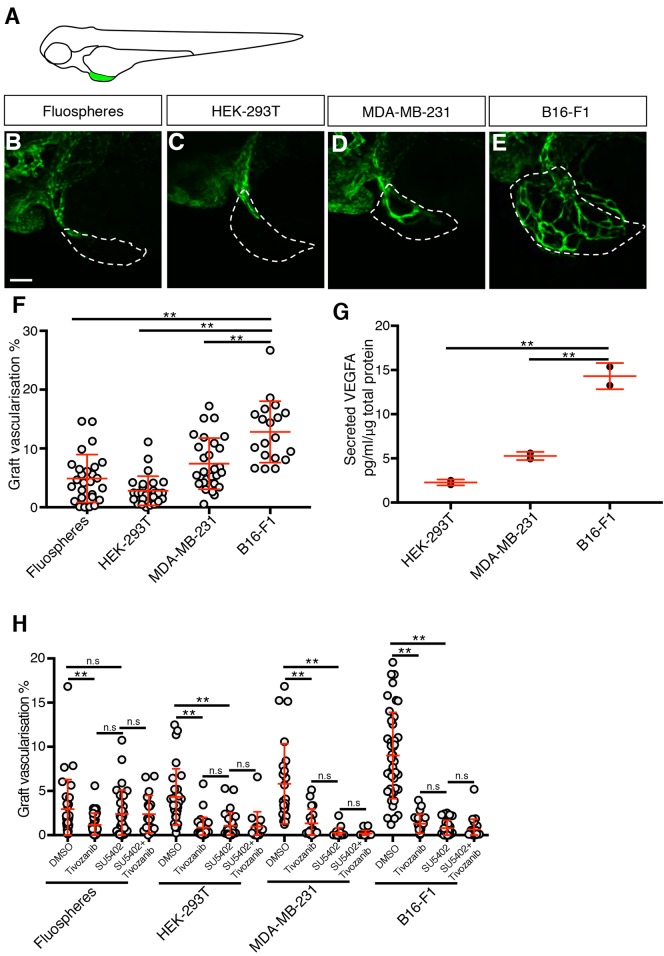
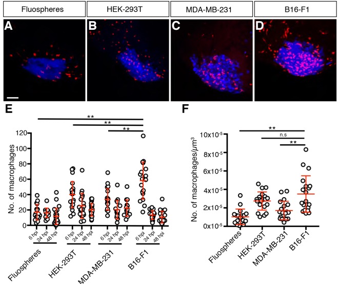
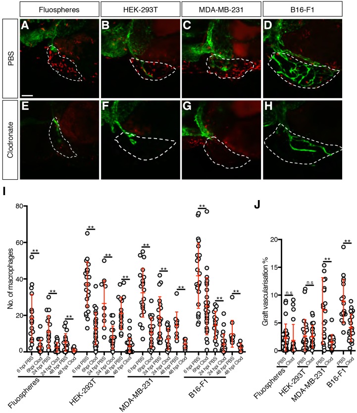
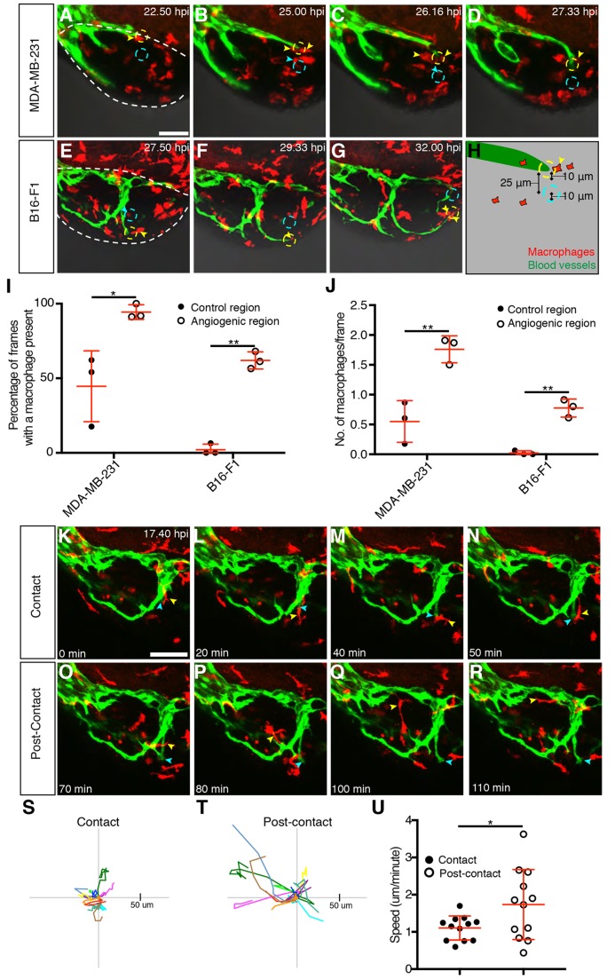
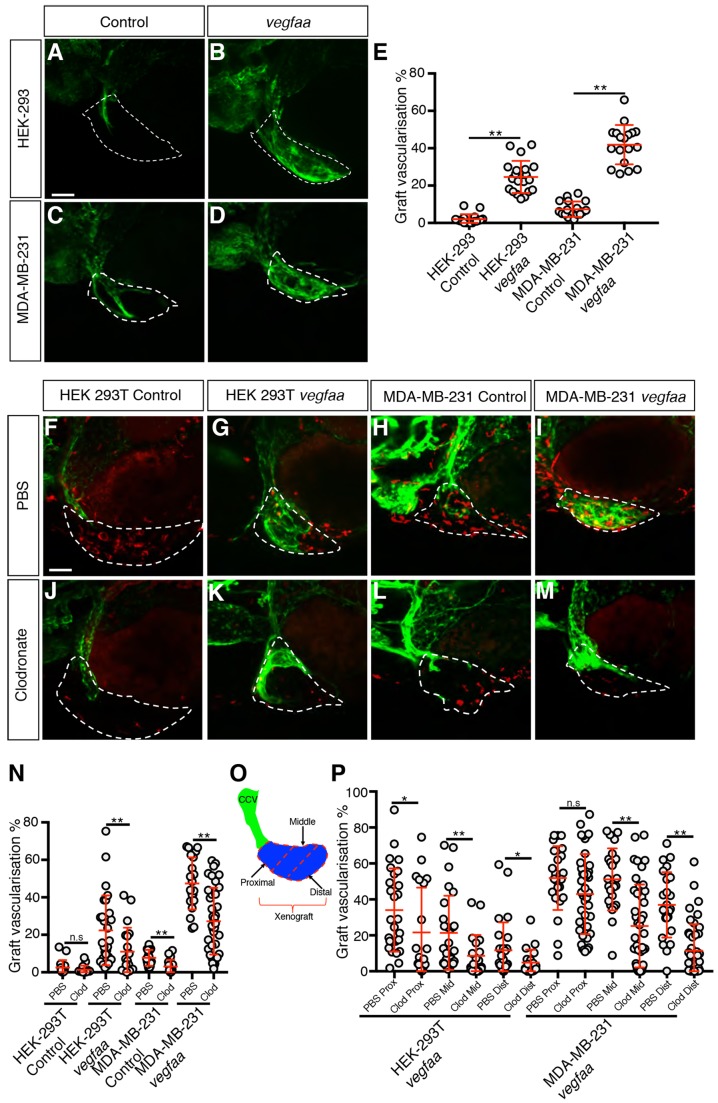
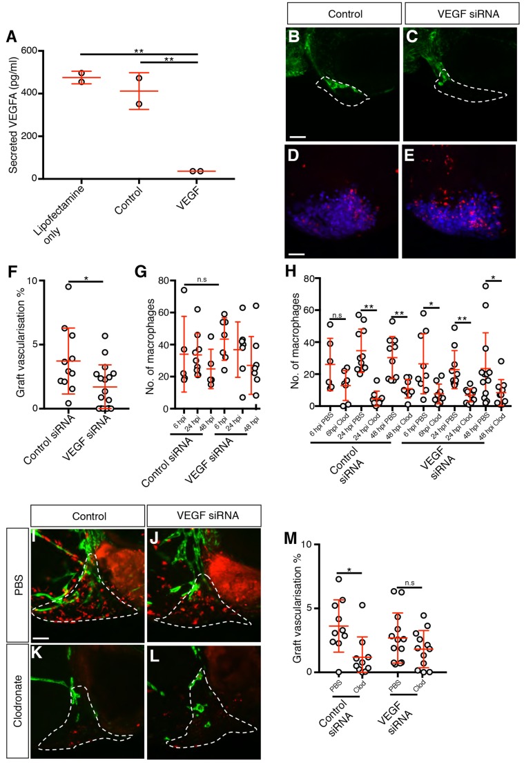
Tumour angiogenesis has long been a focus of anti-cancer therapy; however, anti-angiogenic cancer treatment strategies have had limited clinical success. Tumour-associated myeloid cells are believed to play a role in the resistance of cancer towards anti-angiogenesis therapy, but the mechanisms by which they do this are unclear. An embryonic zebrafish xenograft model has been developed to investigate the mechanisms of tumour angiogenesis and as an assay to screen anti-angiogenic compounds. In this study, we used cell ablation techniques to remove either macrophages or neutrophils and assessed their contribution towards zebrafish xenograft angiogenesis by quantitating levels of graft vascularisation. The ablation of macrophages, but not neutrophils, caused a strong reduction in tumour xenograft vascularisation and time-lapse imaging demonstrated that tumour xenograft macrophages directly associated with the migrating tip of developing tumour blood vessels. Finally, we found that, although macrophages are required for vascularisation in xenografts that either secrete VEGFA or overexpress zebrafish vegfaa, they are not required for the vascularisation of grafts with low levels of VEGFA, suggesting that zebrafish macrophages can enhance Vegfa-driven tumour angiogenesis. The importance of macrophages to this angiogenic response suggests that this model could be used to further investigate the interplay between myeloid cells and tumour vascularisation.

Keywords: Angiogenesis; Macrophage; Tumour; Xenograft; Zebrafish.


Conflict of interest statement

Competing interestsThe authors declare no competing or financial interests.

Figures

Fig. 1.
Fig. 1.
Graft implantation induces an angiogenic response. (A) Schematic indicating the location of the implanted graft (green) in the perivitelline space of a zebrafish embryo. (B-E) Confocal images of kdrl:EGFP-expressing blood vessels (green) in zebrafish embryo grafts (white dashed line) at 2 dpi. (F) Quantitation of graft vascularisation at 2 dpi, n>18. (G) Quantitation of secreted VEGFA levels, n=2. (H) Quantitation of graft vascularisation in embryos incubated in either 0.5% DMSO, 50 nM Tivozanib, 200 nM SU5402 or 50 nM Tivozanib+200 nM SU5402, n>14. Error bars represent s.d. n.s, P>0.05; **P<0.01 by one-way ANOVA. Scale bar: 50 µm.
Fig. 2.
Fig. 2.
Macrophages are recruited to grafts. (A-D) Confocal images of mpeg1:mCherry-expressing macrophages (red) in zebrafish embryo grafts (blue) at 6 hpi. (E) Quantitation of graft-associated macrophages at 6, 24 and 48 hpi, n>14. (F) Quantitation of graft-associated macrophages normalised for graft volume at 6 hpi, n>16. Error bars represent s.d. n.s, P>0.05; **P<0.01 by one-way ANOVA. Scale bar: 50 µm.
Fig. 3.
Fig. 3.
Macrophages contribute to tumour xenograft vascularisation. (A-H) Confocal images of mpeg1:mCherry-expressing macrophages (red) and kdrl:EGFP-expressing blood vessels (green) in zebrafish embryo grafts (white dashed line) at 2 dpi that have been injected with either PBS-containing liposomes (A-D) or clodronate-containing liposomes (E-H). (I) Quantitation of graft-associated macrophages at 6, 24 and 48 hpi with either PBS-containing or clodronate-containing liposomes, n>10. (J) Quantitation of graft vascularisation at 2 dpi, n>16. Error bars represent s.d. n.s, P>0.05; **P<0.01 by t-test. Scale bar: 50 µm.
Fig. 4.
Fig. 4.
Macrophages associate with developing xenograft vessels. (A-G) Still images showing an MDA-MB-231 xenograft (A-D, Movie 3) or a B16-F1 xenograft (E-G, Movie 4) in an embryo with mpeg1:mCherry-expressing macrophages (red) and kdrl:EGFP-expressing blood vessels (green). Individual macrophages associated with the angiogenic region (yellow dashed circle) are indicated with yellow arrowheads, while macrophages associated with the control region (cyan dashed circle) are indicated with cyan arrowheads. The tumour region is outlined by a white dashed line in A and E. (H) Schematic demonstrating the positioning of the 10-μm angiogenic region (dashed yellow outline) at the tip of the blood vessel (green) and the control 10-μm control region (dashed cyan outline). (I) Quantitation of the percentage of frames during which a macrophage was observed at either an angiogenic region or a control region in MDA-MB-231 and B16-F1 xenografts, n=3. (J) Mean number of macrophages observed during each frame, in either the angiogenic region or the control region, in MDA-MB-231 and B16 F1 xenografts, n=3. (K-R) Still images showing a B16-F1 xenograft (Movie 4) in an embryo with mpeg1:mCherry-expressing macrophages (red) and kdrl:EGFP-expressing blood vessels (green). An individual macrophage (yellow arrowhead) is tracked for 1 h 10 min during (K-N) and after (O-R) associating with the distal tip of a growing vessel (indicated by a cyan arrowhead). (S,T) Macrophage migration tracks of 12 macrophages during their period of contact with the tip of a growing vessel (S) and of the same macrophages once they leave the vessel tip (T). Each macrophage is depicted with the same colour in both S and T and they were tracked for identical periods of time during contact and post-contact. (U) Quantitation of macrophage migration speed during and after the period of contact with the tip of a growing vessel, n=12. Error bars represent s.d. *P<0.05, **P<0.01 by t-test. Scale bars: 50 µm.
Fig. 5.
Fig. 5.
Macrophages are required for effective vegfaa-driven angiogenesis. (A-D) Confocal images taken at 2 dpi of kdrl:EGFP-expressing vessels (green) in zebrafish embryos implanted with either HEK-293T (A,B) or MDA-MB-231 (C,D) xenografts (white dashed line) transfected with either a control expression vector (A,C) or a vegfaa-expression vector (B,D). (E) Quantitation of graft vascularisation at 2 dpi, n>17. (F-M) Confocal images taken at 2 dpi of mpeg1:mCherry-expressing macrophages (red) and kdrl:EGFP-expressing blood vessels (green) in zebrafish embryos implanted with either HEK-293T or MDA-MB-231 xenografts (white dashed lines) transfected with either a control expression vector (F,H,J,L) or a vegfaa-expression vector (G,I,K,M) that have been injected with either PBS-containing liposomes (F-I) or clodronate-containing liposomes (J-M). (N) Quantitation of graft vascularisation at 2 dpi in embryos injected with either PBS-containing or clodronate-containing liposomes, n>16. (O) Schematic demonstrating how the proximal, middle and distal sections (marked by dashed red lines) of the xenograft (blue) were determined by their location with respect to the CCV (green). (P) Quantitation of vascularisation at 2 dpi in the proximal, middle and distal regions of vegfaa-expressing HEK-293T or MDA-MB-231 xenografts implanted into embryos injected with either PBS-containing or clodronate-containing liposomes, n>19. Error bars represent s.d. n.s, P>0.05; *P<0.05, **P<0.01 by t-test. Scale bars: 50 µm.
Fig. 6.
Fig. 6.
Macrophages are not required for vascularisation in MDA-MB-231 xenografts depleted of VEGFA. (A) Quantitation of secreted VEGFA levels in 2×10⁵ siRNA-treated cells, n=2. (B,C) Confocal images taken at 2 dpi of kdrl:EGFP-expressing vessels (green) in zebrafish embryos implanted with MDA-MB-231 xenografts (white dashed line) transfected with either control (B) or VEGFA siRNA (C). (D,E) Confocal images taken at 6 hpi of mpeg1:mCherry-expressing macrophages (red) and MDA-MB-231 xenografts (blue). (F,G) Quantitation of graft vascularisation at 2 dpi, n>10 (F), and of graft-associated macrophages at 6, 24 and 48 hpi, n>4 (G). (H) Quantitation of graft-associated macrophages at 6, 24 and 48 hpi, in embryos injected with either PBS-containing or clodronate-containing liposomes, n>5. (I-L) Confocal images taken at 2 dpi of mpeg1:mCherry-expressing macrophages (red) and kdrl:EGFP-expressing blood vessels (green) in zebrafish embryos implanted with MDA-MB-231 xenografts (white dashed lines) transfected with either control (I,K) or VEGFA (J,L) siRNA that have been injected with either PBS-containing liposomes (I,J) or clodronate-containing liposomes (K,L). (M) Quantitation of graft vascularisation at 2 dpi in embryos injected with either PBS-containing or clodronate-containing liposomes, n>9. Error bars represent s.d. n.s, P>0.05; *P<0.05; **P<0.01 by either one-way ANOVA (A) or t-test (F-H,M). Scale bars: 50 µm.

References

    1. Ablain J., Durand E. M., Yang S., Zhou Y. and Zon L. I. (2015). A CRISPR/Cas9 vector system for tissue-specific gene disruption in zebrafish. Dev. Cell 32, 756-764. 10.1016/j.devcel.2015.01.032 - DOI - PMC - PubMed
    1. Albini A., Bruno A., Noonan D. M. and Mortara L. (2018). Contribution to tumor angiogenesis from innate immune cells within the tumor microenvironment: implications for immunotherapy. Front. Immunol. 9, 527 10.3389/fimmu.2018.00527 - DOI - PMC - PubMed
    1. Ardi V. C., Van den Steen P. E., Opdenakker G., Schweighofer B., Deryugina E. I. and Quigley J. P. (2009). Neutrophil MMP-9 proenzyme, unencumbered by TIMP-1, undergoes efficient activation in vivo and catalytically induces angiogenesis via a basic fibroblast growth factor (FGF-2)/FGFR-2 pathway. J. Biol. Chem. 284, 25854-25866. 10.1074/jbc.M109.033472 - DOI - PMC - PubMed
    1. Asahara T., Murohara T., Sullivan A., Silver M., van der Zee R., Li T., Witzenbichler B., Schatteman G. and Isner J. M. (1997). Isolation of putative progenitor endothelial cells for angiogenesis. Science 275, 964-967. 10.1126/science.275.5302.964 - DOI - PubMed
    1. Astin J. W., Haggerty M. J. L., Okuda K. S., Le Guen L., Misa J. P., Tromp A., Hogan B. M., Crosier K. E. and Crosier P. S. (2014). Vegfd can compensate for loss of Vegfc in zebrafish facial lymphatic sprouting. Development 141, 2680 10.1242/dev.106591 - DOI - PubMed

Publication types

MeSH terms

Substances