

Critical Role of Macrophage FcγR Signaling and Reactive Oxygen Species in Alloantibody-Mediated Hepatocyte Rejection
- PMID: 30397035
- PMCID: PMC6289737
- DOI: 10.4049/jimmunol.1800333
Critical Role of Macrophage FcγR Signaling and Reactive Oxygen Species in Alloantibody-Mediated Hepatocyte Rejection
Abstract
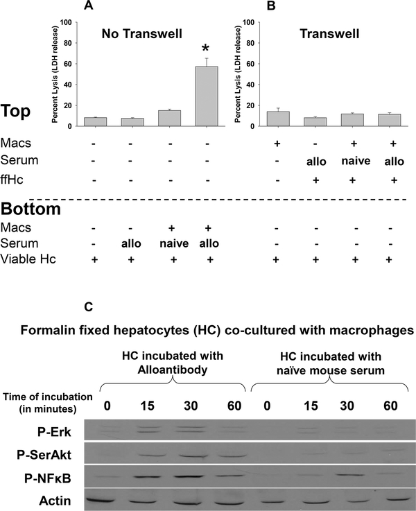
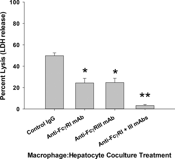
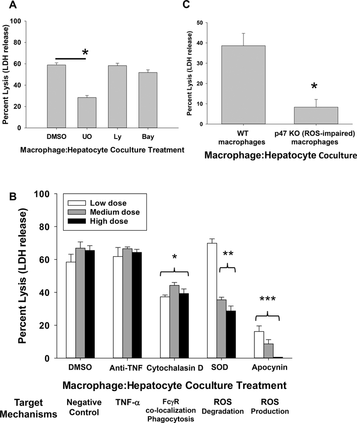
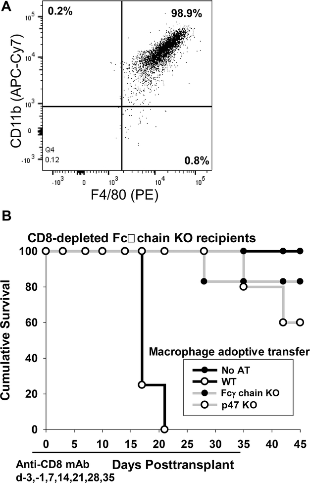
Humoral alloimmunity negatively impacts both short- and long-term cell and solid organ transplant survival. We previously reported that alloantibody-mediated rejection of transplanted hepatocytes is critically dependent on host macrophages. However, the effector mechanism(s) of macrophage-mediated injury to allogeneic liver parenchymal cells is not known. We hypothesized that macrophage-mediated destruction of allogeneic hepatocytes occurs by cell-cell interactions requiring FcγRs. To examine this, alloantibody-dependent hepatocyte rejection in CD8-depleted wild-type (WT) and Fcγ-chain knockout (KO; lacking all functional FcγR) transplant recipients was evaluated. Alloantibody-mediated hepatocellular allograft rejection was abrogated in recipients lacking FcγR compared with WT recipients. We also investigated anti-FcγRI mAb, anti-FcγRIII mAb, and inhibitors of intracellular signaling (to block phagocytosis, cytokines, and reactive oxygen species [ROS]) in an in vitro alloantibody-dependent, macrophage-mediated hepatocytoxicity assay. Results showed that in vitro alloantibody-dependent, macrophage-mediated hepatocytotoxicity was critically dependent on FcγRs and ROS. The adoptive transfer of WT macrophages into CD8-depleted FcγR-deficient recipients was sufficient to induce alloantibody-mediated rejection, whereas adoptive transfer of macrophages from Fcγ-chain KO mice or ROS-deficient (p47 KO) macrophages was not. These results provide the first evidence, to our knowledge, that alloantibody-dependent hepatocellular allograft rejection is mediated by host macrophages through FcγR signaling and ROS cytotoxic effector mechanisms. These results support the investigation of novel immunotherapeutic strategies targeting macrophages, FcγRs, and/or downstream molecules, including ROS, to inhibit humoral immune damage of transplanted hepatocytes and perhaps other cell and solid organ transplants.
Copyright © 2018 by The American Association of Immunologists, Inc.
Figures

Similar articles
-
CD4+ T-cell-dependent immune damage of liver parenchymal cells is mediated by alloantibody.Transplantation. 2005 Aug 27;80(4):514-21. doi: 10.1097/01.tp.0000168342.57948.68. Transplantation. 2005. PMID: 16123727
-
Critical role of effector macrophages in mediating CD4-dependent alloimmune injury of transplanted liver parenchymal cells.J Immunol. 2008 Jul 15;181(2):1224-31. doi: 10.4049/jimmunol.181.2.1224. J Immunol. 2008. PMID: 18606676 Free PMC article.
-
mTOR Inhibition Suppresses Posttransplant Alloantibody Production Through Direct Inhibition of Alloprimed B Cells and Sparing of CD8+ Antibody-Suppressing T cells.Transplantation. 2016 Sep;100(9):1898-906. doi: 10.1097/TP.0000000000001291. Transplantation. 2016. PMID: 27362313 Free PMC article.
-
Unusual patterns of alloimmunity evoked by allogeneic liver parenchymal cells.Immunol Rev. 2000 Apr;174:260-79. doi: 10.1034/j.1600-0528.2002.017409.x. Immunol Rev. 2000. PMID: 10807522 Review.
-
B cells: a rational target in alloantibody-mediated solid organ transplantation rejection.Clin Transplant. 2006 Jan-Feb;20(1):48-54. doi: 10.1111/j.1399-0012.2005.00439.x. Clin Transplant. 2006. PMID: 16556153 Review.
Cited by
-
Antibody-suppressor CD8+ T Cells Require CXCR5.Transplantation. 2019 Sep;103(9):1809-1820. doi: 10.1097/TP.0000000000002683. Transplantation. 2019. PMID: 30830040 Free PMC article.
-
Gut redox and microbiome: charting the roadmap to T-cell regulation.Front Immunol. 2024 Aug 21;15:1387903. doi: 10.3389/fimmu.2024.1387903. eCollection 2024. Front Immunol. 2024. PMID: 39234241 Free PMC article. Review.
-
Microvascular Inflammation of the Renal Allograft: A Reappraisal of the Underlying Mechanisms.Front Immunol. 2022 Mar 22;13:864730. doi: 10.3389/fimmu.2022.864730. eCollection 2022. Front Immunol. 2022. PMID: 35392097 Free PMC article. Review.
-
Macrophages and hepatocellular carcinoma.Cell Biosci. 2019 Sep 26;9:79. doi: 10.1186/s13578-019-0342-7. eCollection 2019. Cell Biosci. 2019. PMID: 31572568 Free PMC article. Review.
-
Reactive Oxygen Species in Autoimmune Cells: Function, Differentiation, and Metabolism.Front Immunol. 2021 Feb 25;12:635021. doi: 10.3389/fimmu.2021.635021. eCollection 2021. Front Immunol. 2021. PMID: 33717180 Free PMC article. Review.
References
-
- Colvin RB, and Smith RN 2005. Antibody-mediated organ-allograft rejection. Nat. Rev. Immunol 5: 807–817. - PubMed
-
- Lorenz M, Regele H, Schillinger M, Exner M, Rasoul-Rockenschaub S, Wahrmann M, Kletzmayr J, Silberhumer G, Horl WH, and Bohmig GA 2004. Risk factors for capillary C4d deposition in kidney allografts: evaluation of a large study cohort. Transplantation. 78: 447–452. - PubMed
-
- Ryan EA, Paty BW, Senior PA, Bigam D, Alfadhli E, Kneteman NM, Lakey JR, and Shapiro AM 2005. Five-year follow-up after clinical islet transplantation. Diabetes. 54: 2060–2069. - PubMed
-
- Jorns C, Nowak G, Nemeth A, Zemack H, Mork LM, Johansson H, Gramignoli R, Watanabe M, Karadagi A, Alheim M, Hauzenberger D, van Dijk R, Bosma PJ, Ebbesen F, Szakos A, Fischler B, Strom S, Ellis E, and Ericzon BG 2016. De Novo Donor-Specific HLA Antibody Formation in Two Patients With Crigler-Najjar Syndrome Type I Following Human Hepatocyte Transplantation With Partial Hepatectomy Preconditioning. Am. J. Transplant 16: 1021–1030. - PMC - PubMed
-
- Colvin RB 2007. Antibody-mediated renal allograft rejection: diagnosis and pathogenesis. J. Am. Soc. Nephrol 18: 1046–1056. - PubMed
Publication types
MeSH terms
Substances
Grants and funding
LinkOut - more resources
Full Text Sources
Molecular Biology Databases
Research Materials
