. 2019 Jan 3;39(2):e00332-18.
doi: 10.1128/MCB.00332-18. Print 2019 Jan 15.

Interleukin-8 Activates Breast Cancer-Associated Adipocytes and Promotes Their Angiogenesis- and Tumorigenesis-Promoting Effects

Affiliations

Interleukin-8 Activates Breast Cancer-Associated Adipocytes and Promotes Their Angiogenesis- and Tumorigenesis-Promoting Effects

Huda H Al-Khalaf et al. Mol Cell Biol. .

Abstract

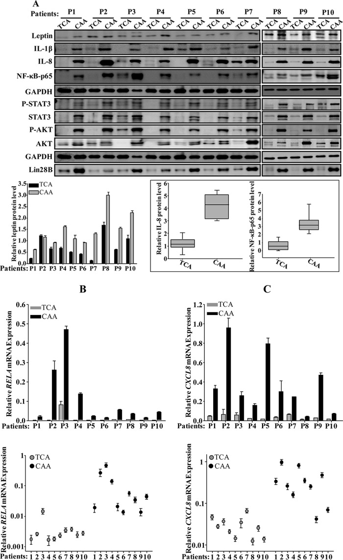
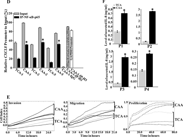
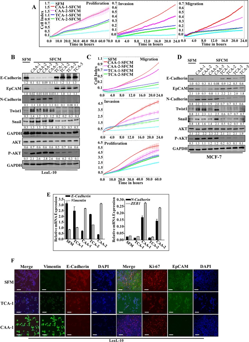
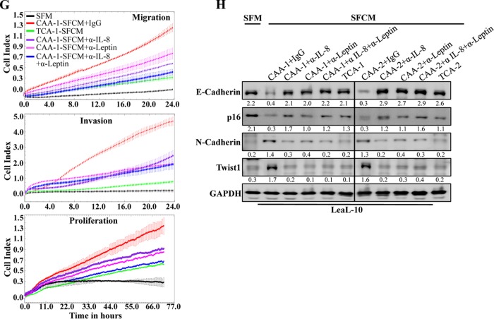
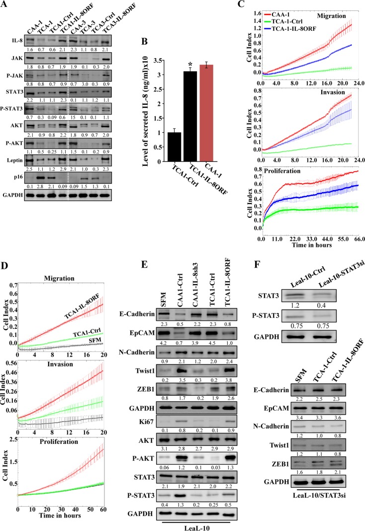
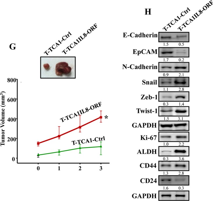
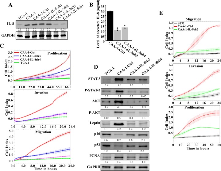
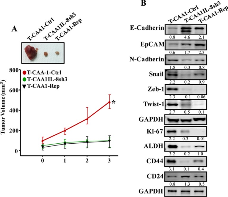
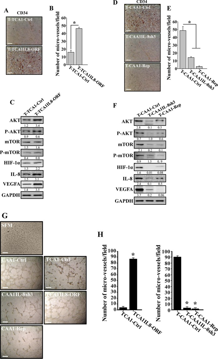
Increasing evidence supports the critical role of active stromal adipocytes in breast cancer development and spread. However, the mediators and the mechanisms of action are still elusive. We show here that cancer-associated adipocytes (CAAs) isolated from 10 invasive breast carcinomas are proinflammatory and exhibit active phenotypes, including higher proliferative, invasive, and migratory capacities compared to their adjacent tumor-counterpart adipocytes (TCAs). Furthermore, all CAAs secreted higher level of interleukin-8 (IL-8), which is critical in mediating the paracrine procarcinogenic effects of these cells. Importantly, ectopic expression of IL-8 in TCA cells activated them and enhanced their procarcinogenic effects both in vitro, in a STAT3-dependent manner, and in vivo In contrast, inhibition of the IL-8 signaling using specific short hairpin RNA, anti-IL-8 antibody, or reparixin suppressed the active features of CAAs, including their non-cell-autonomous tumor-promoting activities both on breast luminal cells and in orthotopic tumor xenografts in mice. IL-8 played also an important role in enhancing the proangiogenic effects of breast adipocytes. These results provide clear indication that IL-8 plays key roles in the activation of breast CAAs and acts as a major mediator for their paracrine protumorigenic effects. Thus, targeting CAAs by inhibiting the IL-8 pathway could have great therapeutic value.

Keywords: IL-8; STAT3; adipocytes; breast cancer; reparixin.


Figures

FIG 1
FIG 1
Mature breast cancer-associated adipocytes are active. (A) Whole-cell lysates were prepared from the indicated mature adipocytes and utilized for immunoblotting with antibodies against the indicated proteins. (Lower panel, left) Quantification of leptin expression levels. (Right) Box plot showing IL-8 and NF-κB (p65) expression levels in the indicated types of breast adipocytes. (B and C) Total RNA was prepared from the indicated adipocytes and utilized to assess the levels of the CXCL8 and RELA mRNAs by qRT-PCR using GAPDH as a reference gene. Error bars represent means ± the SD, and values reflect three independent experiments. Upper panels show the difference in the expression of individual transcripts between CAA and TCA cells isolated from the same patients, as indicated. Lower panels show the difference between all the CAAs compared to their corresponding TCAs. (D) ChIP assay. Chromatin was purified from the indicated adipocytes and then immunoprecipitated with the indicated antibodies. Subsequently, the CXCL8 promoter bearing the NF-κB-p65 binding site was amplified by qPCR using specific primers. The GAPDH promoter was used as an unlinked locus control, and the abundance of the promoter was plotted relative to GAPDH after normalization of each sample against its own input. These experiments were performed three times, and error bars represent means ± the SD. *, P ≤ 4.1 × 10−5. (E) The indicated CAAs and TCAs were seeded in SFM in the upper wells of the CIM plates, and the migration/invasion abilities were assessed by the RTCA-xCELLigence system. For proliferation, adipocytes were seeded in complete medium in the E-plate, and the proliferation rate was assessed by the RTCA-xCELLigence system. (F) SFCM from four subtypes of CAAs and their corresponding TCA cells were utilized to assess the secreted levels of IL-8 by ELISA. Error bars represent means ± the SD; values were determined from three different experiments. *, P ≤ 0.04.
FIG 1
FIG 1
Mature breast cancer-associated adipocytes are active. (A) Whole-cell lysates were prepared from the indicated mature adipocytes and utilized for immunoblotting with antibodies against the indicated proteins. (Lower panel, left) Quantification of leptin expression levels. (Right) Box plot showing IL-8 and NF-κB (p65) expression levels in the indicated types of breast adipocytes. (B and C) Total RNA was prepared from the indicated adipocytes and utilized to assess the levels of the CXCL8 and RELA mRNAs by qRT-PCR using GAPDH as a reference gene. Error bars represent means ± the SD, and values reflect three independent experiments. Upper panels show the difference in the expression of individual transcripts between CAA and TCA cells isolated from the same patients, as indicated. Lower panels show the difference between all the CAAs compared to their corresponding TCAs. (D) ChIP assay. Chromatin was purified from the indicated adipocytes and then immunoprecipitated with the indicated antibodies. Subsequently, the CXCL8 promoter bearing the NF-κB-p65 binding site was amplified by qPCR using specific primers. The GAPDH promoter was used as an unlinked locus control, and the abundance of the promoter was plotted relative to GAPDH after normalization of each sample against its own input. These experiments were performed three times, and error bars represent means ± the SD. *, P ≤ 4.1 × 10−5. (E) The indicated CAAs and TCAs were seeded in SFM in the upper wells of the CIM plates, and the migration/invasion abilities were assessed by the RTCA-xCELLigence system. For proliferation, adipocytes were seeded in complete medium in the E-plate, and the proliferation rate was assessed by the RTCA-xCELLigence system. (F) SFCM from four subtypes of CAAs and their corresponding TCA cells were utilized to assess the secreted levels of IL-8 by ELISA. Error bars represent means ± the SD; values were determined from three different experiments. *, P ≤ 0.04.
FIG 2
FIG 2
CAAs promote EMT in normal breast luminal cells in an IL-8-dependent manner. LeaL-10 cells were exposed to SFM or SFCM from four CAAs and their four corresponding TCA cells for 24 h. (A and C) Migration/invasion and proliferation capabilities were assessed for the indicated pairs. The graphs are representative of different experiments. (B and D) Cell lysates were prepared from the indicated cells and used for immunoblotting. (E) LeaL-10 cells were grown in 3D cultures and then either exposed to two CAAs and their two corresponding TCAs or SFM. Total RNA was prepared and used to assess the level of the indicated transcripts by qRT-PCR. Error bars represent means ± the SD, and values were drawn from three different experiments. (F) LeaL-10 cells were treated as for panel E, cytospun, and used for immunofluorescence. Scale bars, 50 µm. (G) LeaL-10 cells were exposed to SFM or SFCM from CAA1 containing either anti-IL-8, antileptin, anti-IL-8 plus antileptin, or anti-IgG antibodies and then utilized to assess migration/invasion and proliferation capabilities. The graphs are representative of different experiments. (H) LeaL-10 cells were treated as indicated, and then whole-cell lysates were prepared and used to assess the levels of the indicated proteins by immunoblotting. For the immunoblots, the numbers below the bands indicate the corresponding protein expression levels relative to GAPDH. The levels of phosphorylated proteins were normalized against the total amounts of the respective nonphosphorylated proteins.
FIG 2
FIG 2
CAAs promote EMT in normal breast luminal cells in an IL-8-dependent manner. LeaL-10 cells were exposed to SFM or SFCM from four CAAs and their four corresponding TCA cells for 24 h. (A and C) Migration/invasion and proliferation capabilities were assessed for the indicated pairs. The graphs are representative of different experiments. (B and D) Cell lysates were prepared from the indicated cells and used for immunoblotting. (E) LeaL-10 cells were grown in 3D cultures and then either exposed to two CAAs and their two corresponding TCAs or SFM. Total RNA was prepared and used to assess the level of the indicated transcripts by qRT-PCR. Error bars represent means ± the SD, and values were drawn from three different experiments. (F) LeaL-10 cells were treated as for panel E, cytospun, and used for immunofluorescence. Scale bars, 50 µm. (G) LeaL-10 cells were exposed to SFM or SFCM from CAA1 containing either anti-IL-8, antileptin, anti-IL-8 plus antileptin, or anti-IgG antibodies and then utilized to assess migration/invasion and proliferation capabilities. The graphs are representative of different experiments. (H) LeaL-10 cells were treated as indicated, and then whole-cell lysates were prepared and used to assess the levels of the indicated proteins by immunoblotting. For the immunoblots, the numbers below the bands indicate the corresponding protein expression levels relative to GAPDH. The levels of phosphorylated proteins were normalized against the total amounts of the respective nonphosphorylated proteins.
FIG 3
FIG 3
IL-8 upregulation activates breast stromal adipocytes. (A) TCA1 and TCA3 cells were transfected with a plasmid bearing IL-8 ORF (TCA1-IL8ORF and TCA3-IL8ORF, respectively; an empty plasmid was used as the control, TCA1-Ctrl and TCA3-Ctrl, respectively), and cell lysates were prepared and utilized for immunoblotting, using CAA1 and CAA3 as controls, respectively. (B) SFCM were collected from the indicated cells and utilized to assess the levels of secreted IL-8 by ELISA. Error bars represent the means ± the SD of three different experiments. *, P = 0.018. (C) Same as for Fig. 1F. (D and E) LeaL-10 cells were exposed to SFM, SFCM from TCA1-IL8ORF, or TCA1-Ctr for 24 h and then were utilized to assess migration/invasion and proliferation capabilities (D) (the graphs are representative of different experiments) and to prepare whole-cell lysates to assess the levels of the indicated proteins by immunoblotting (E). (F) LeaL-10 cells were transfected with STAT3 siRNA and then exposed to SFM, SFCM from TCA1-IL8ORF, or TCA1-Ctrl for 24 h and were then utilized to prepare whole-cell lysates to assess the levels of the indicated proteins by immunoblotting. (G) Breast cancer xenografts were created by coinjecting MDA-MB-231 cells with TCA1-IL8ORF or TCA1-Ctrl cells (n = 4/each group) under the nipples of nude mice. (Upper panel) Representative tumor size. (Lower panel) Graph showing time-dependent tumor growth. Error bars represent the means ± the SD, with values from four mice. *, P= 0.014. (H) Tumors were excised, and whole-tissue lysates were prepared and utilized for immunoblotting. For the immunoblots, the numbers below the bands indicate the corresponding protein expression levels relative to GAPDH. The levels of phosphorylated proteins were normalized against the total amounts of the respective nonphosphorylated proteins.
FIG 3
FIG 3
IL-8 upregulation activates breast stromal adipocytes. (A) TCA1 and TCA3 cells were transfected with a plasmid bearing IL-8 ORF (TCA1-IL8ORF and TCA3-IL8ORF, respectively; an empty plasmid was used as the control, TCA1-Ctrl and TCA3-Ctrl, respectively), and cell lysates were prepared and utilized for immunoblotting, using CAA1 and CAA3 as controls, respectively. (B) SFCM were collected from the indicated cells and utilized to assess the levels of secreted IL-8 by ELISA. Error bars represent the means ± the SD of three different experiments. *, P = 0.018. (C) Same as for Fig. 1F. (D and E) LeaL-10 cells were exposed to SFM, SFCM from TCA1-IL8ORF, or TCA1-Ctr for 24 h and then were utilized to assess migration/invasion and proliferation capabilities (D) (the graphs are representative of different experiments) and to prepare whole-cell lysates to assess the levels of the indicated proteins by immunoblotting (E). (F) LeaL-10 cells were transfected with STAT3 siRNA and then exposed to SFM, SFCM from TCA1-IL8ORF, or TCA1-Ctrl for 24 h and were then utilized to prepare whole-cell lysates to assess the levels of the indicated proteins by immunoblotting. (G) Breast cancer xenografts were created by coinjecting MDA-MB-231 cells with TCA1-IL8ORF or TCA1-Ctrl cells (n = 4/each group) under the nipples of nude mice. (Upper panel) Representative tumor size. (Lower panel) Graph showing time-dependent tumor growth. Error bars represent the means ± the SD, with values from four mice. *, P= 0.014. (H) Tumors were excised, and whole-tissue lysates were prepared and utilized for immunoblotting. For the immunoblots, the numbers below the bands indicate the corresponding protein expression levels relative to GAPDH. The levels of phosphorylated proteins were normalized against the total amounts of the respective nonphosphorylated proteins.
FIG 4
FIG 4
IL-8 downregulation suppresses the activity features of CAA. (A) CAA1 cells were separately transfected with four different specific IL-8-shRNAs (CAA1-IL8sh1, CAA1-IL8sh2, CAA1-IL8sh3, and CAA1-IL8sh4), as well as a control plasmid bearing a scrambled sequence (CAA1-Ctrl), and then cell lysates were prepared and used for immunoblotting. (B) SFCM collected from the indicated cells were utilized to assess the levels of secreted IL-8 by ELISA. Error bars represent means ± the SD, and the values represent three independent experiments. *, P ≤ 0.000246. (C) Same as for Fig. 1F. (D) Cell lysates were prepared from the indicated cells and used for immunoblotting analysis. For the immunoblots, the numbers below the bands indicate the corresponding protein expression levels relative to GAPDH. The levels of phosphorylated proteins were normalized against the total amounts of the respective nonphosphorylated proteins. (E) LeaL-10 cells were exposed to SFM or SFCM from the indicated cells and then were utilized to assess the migration/invasion and proliferation capabilities by the RTCA-xCELLigence system. The graphs are representative of different experiments.
FIG 5
FIG 5
Inhibition of CXCR1/2 persistently suppresses the CAA activity. (A) CAA1 cells were treated with different doses of reparixin for 24 h, and anti-IL-8 neutralizing antibody was used as a positive control. Cell lysates were prepared and utilized to assess the level of the indicated proteins using specific antibodies. The numbers below the bands indicate the corresponding protein expression levels relative to GAPDH. The levels of phosphorylated proteins were normalized against the total amounts of the respective nonphosphorylated proteins. (B) CAA1 cells were treated as described above and seeded in SFM in the upper wells of the CIM plates, and the migration/invasion abilities were assessed. For proliferation, pretreated CAA1 cells were seeded in complete medium in the E-plate, and the proliferation rate was assessed by the RTCA-xCELLigence system. (C) SFCM were collected from the indicated cells (either 24 h posttreatment [24 h] or 72 h postsplit [split]) and then utilized to assess the levels of secreted IL-8 by ELISA. Error bars represent means ± the SD of three different experiments. *, P ≤ 0.018. (D and E) Total RNA was prepared from the indicated adipocytes (either 72 h postsplit [split] or after the fourth split [split-4]) and then utilized to assess the levels of the indicated transcripts by qRT-PCR. Error bars represent means ± the SD, and the values represent three independent experiments. *, P ≤ 0.0015. (F) LeaL-10 cells were exposed to SFM or SFCM from CAA1 cells treated either with reparixin or anti-IL-8 antibody, and total RNA was prepared and used to assess the levels of the indicated transcripts by qRT-PCR. Error bars represent means ± the SD, and values represent three different experiments. *, P ≤ 0.0037.
FIG 6
FIG 6
CAA cells stimulate breast cancer xenograft growth in mice in an IL-8-dependent manner. (A) Breast cancer xenografts were created by coinjecting MDA-MB-231 cells with CAA1-Ctrl, CAA1-IL8sh3 or CAA1 pretreated with reparixin (100 ng) for 24 h (CAA1-Rep) (n = 4/each group) under the nipples of nude mice. (Upper panel) Representative tumor sizes. (Lower panel) Time-dependent tumor growth. Error bars represent means ± the SD, and the values are derived from four mice. *, P = 0.014. (B) Tumors were excised, and whole tissue lysates were prepared and utilized for immunoblotting. The numbers below the bands indicate the corresponding protein expression levels relative to GAPDH.
FIG 7
FIG 7
CAA cells induce angiogenesis in vivo in an IL-8-depednent manner. (A and D) FFPE tissues were prepared from the indicated tumors, and immunohistochemistry was carried out with anti-CD34 antibody. Scale bars, 50 µm. (B and E) Histograms show the average numbers of CD34+ microvessels observed in five different fields from the different tumors. Error bars represent means ± the SD. *, P ≤ 0.0013. (C and F) Same as in Fig. 3H and 6B. The numbers below the bands indicate the corresponding protein expression levels relative to GAPDH. The levels of phosphorylated proteins were normalized against the total amount of the respective nonphosphorylated proteins. (G) SFM or SFCM from the indicated adipocytes were collected and were added independently on HUVEC cells plated on Matrigel (96-well plate), and the differentiation into capillary-like structures was assessed after 5 h of incubation. Representative photographs of HUVEC cavities are shown. Scale bars, 30 µm. (H) A histogram shows the average numbers of microvessels counted in five different fields. Error bars represent means ± the SD. *, P ≤ 1.19 × 10−6.

Similar articles

Cited by

References

    1. Dirat B, Bochet L, Dabek M, Daviaud D, Dauvillier S, Majed B, Wang YY, Meulle A, Salles B, Le Gonidec S, Garrido I, Escourrou G, Valet P, Muller C. 2011. Cancer-associated adipocytes exhibit an activated phenotype and contribute to breast cancer invasion. Cancer Res 71:2455–2465. doi:10.1158/0008-5472.CAN-10-3323. - DOI - PubMed
    1. Wang YY, Attane C, Milhas D, Dirat B, Dauvillier S, Guerard A, Gilhodes J, Lazar I, Alet N, Laurent V, Le Gonidec S, Biard D, Herve C, Bost F, Ren GS, Bono F, Escourrou G, Prentki M, Nieto L, Valet P, Muller C. 2017. Mammary adipocytes stimulate breast cancer invasion through metabolic remodeling of tumor cells. JCI Insight 2:e87489. doi:10.1172/jci.insight.87489. - DOI - PMC - PubMed
    1. Duong MN, Geneste A, Fallone F, Li X, Dumontet C, Muller C. 2017. The fat and the bad: mature adipocytes, key actors in tumor progression and resistance. Oncotarget 8:57622–57641. doi:10.18632/oncotarget.18038. - DOI - PMC - PubMed
    1. Cirillo D, Rachiglio AM, la Montagna R, Giordano A, Normanno N. 2008. Leptin signaling in breast cancer: an overview. J Cell Biochem 105:956–964. doi:10.1002/jcb.21911. - DOI - PubMed
    1. Ando S, Catalano S. 2011. The multifactorial role of leptin in driving the breast cancer microenvironment. Nat Rev Endocrinol 8:263–275. doi:10.1038/nrendo.2011.184. - DOI - PubMed

Publication types

MeSH terms