. 2018 Nov 20;115(47):E11120-E11127.
doi: 10.1073/pnas.1802519115. Epub 2018 Nov 5.

Expression of ABCA4 in the retinal pigment epithelium and its implications for Stargardt macular degeneration

Affiliations

Expression of ABCA4 in the retinal pigment epithelium and its implications for Stargardt macular degeneration

Tamara L Lenis et al. Proc Natl Acad Sci U S A. .

Abstract

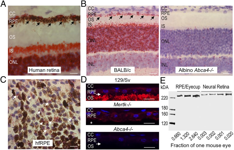
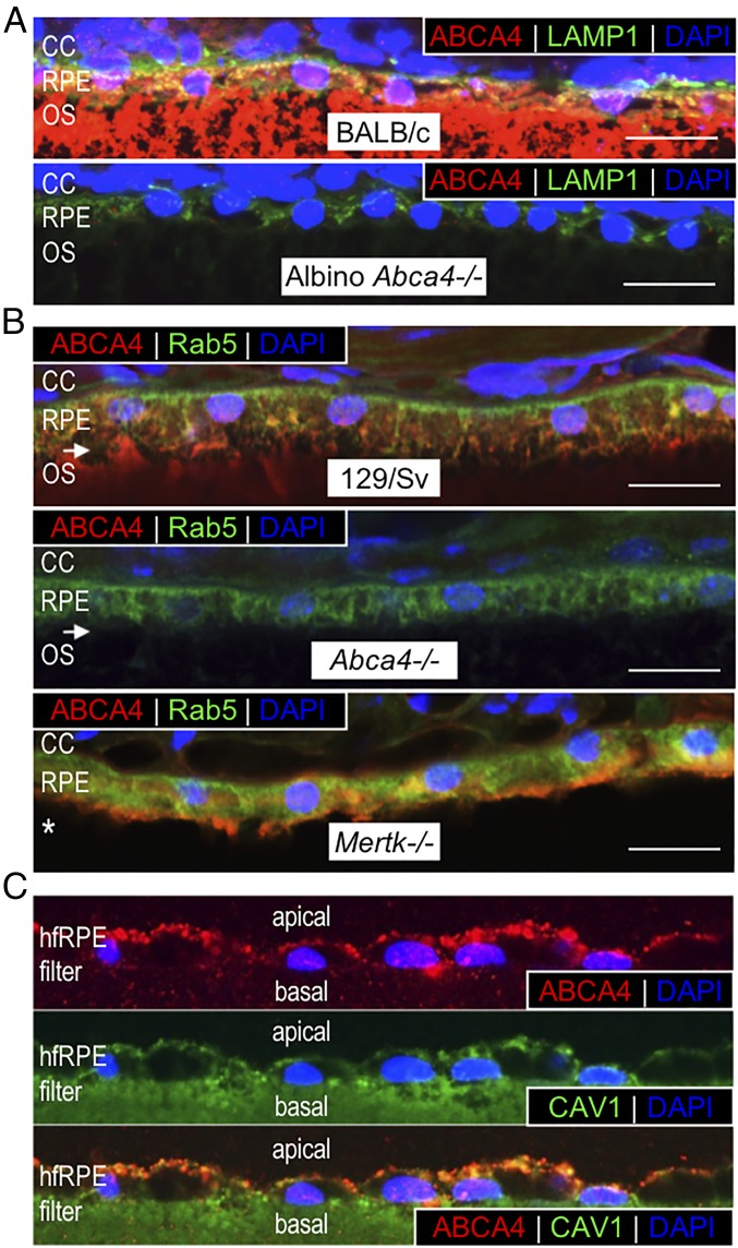
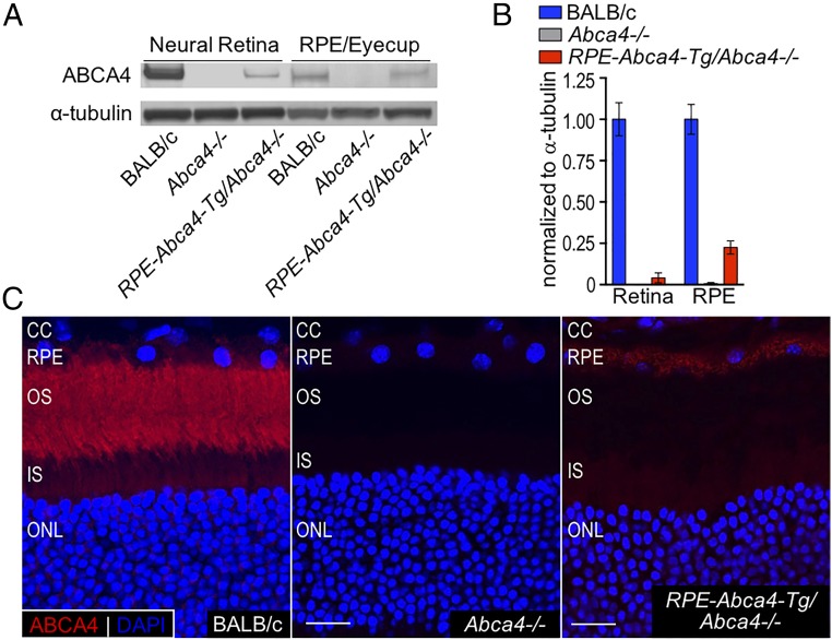
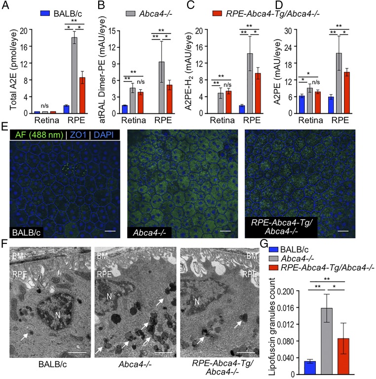
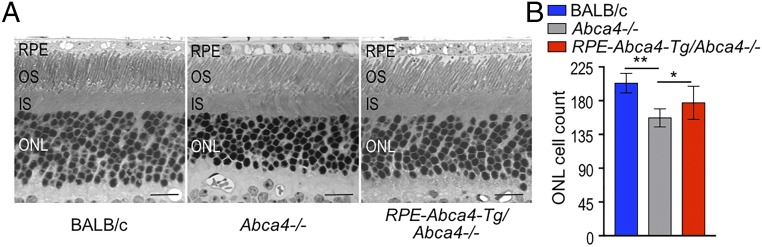
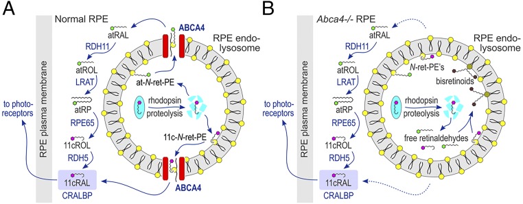
Recessive Stargardt disease (STGD1) is an inherited blinding disorder caused by mutations in the Abca4 gene. ABCA4 is a flippase in photoreceptor outer segments (OS) that translocates retinaldehyde conjugated to phosphatidylethanolamine across OS disc membranes. Loss of ABCA4 in Abca4-/- mice and STGD1 patients causes buildup of lipofuscin in the retinal pigment epithelium (RPE) and degeneration of photoreceptors, leading to blindness. No effective treatment currently exists for STGD1. Here we show by several approaches that ABCA4 is additionally expressed in RPE cells. (i) By in situ hybridization analysis and by RNA-sequencing analysis, we show the Abca4 mRNA is expressed in human and mouse RPE cells. (ii) By quantitative immunoblotting, we show that the level of ABCA4 protein in homogenates of wild-type mouse RPE is about 1% of the level in neural retina homogenates. (iii) ABCA4 immunofluorescence is present in RPE cells of wild-type and Mertk-/- but not Abca4-/- mouse retina sections, where it colocalizes with endolysosomal proteins. To elucidate the role of ABCA4 in RPE cells, we generated a line of genetically modified mice that express ABCA4 in RPE cells but not in photoreceptors. Mice from this line on the Abca4-/- background showed partial rescue of photoreceptor degeneration and decreased lipofuscin accumulation compared with nontransgenic Abca4-/- mice. We propose that ABCA4 functions to recycle retinaldehyde released during proteolysis of rhodopsin in RPE endolysosomes following daily phagocytosis of distal photoreceptor OS. ABCA4 deficiency in the RPE may play a role in the pathogenesis of STGD1.

Keywords: ABCA4; Stargardt disease; bisretinoid; lipofuscin; retinal pigment epithelium.


Conflict of interest statement

The authors declare no conflict of interest.

Figures

Fig. 1.
Fig. 1.
(AC) Abca4 mRNA and protein is expressed in RPE cells. In situ hybridization using the RNAscope assay with an Abca4-specific probe on human cadaveric ocular sections (A), mouse retina sections (B), and hfRPE cells in culture (C). Note the intense chromogenic reactivity (red punctate staining, indicated by the black arrows) for Abca4 mRNA in outer nuclear layer (ONL) and inner segments (IS) of the photoreceptor cells and in RPE cells of human (A) and wild-type BALB/c sections (B, Left). This reactivity is absent in Abca4−/− tissue (B, Right). Red punctate staining (white arrows) corresponding to ABCA4 mRNA is also observed in hfRPE cultured cells (C). CC, choriocapillaris; INL, inner nuclear layer. (Scale bars, 20 μm.) (D) ABCA4 immunohistochemistry (red fluorescence) on retina sections from pigmented wild-type (129/Sv), Mertk−/−, and Abca4−/− mice. Note that ABCA4 immunoreactivity is seen in the RPE and OS of 129/Sv mice and in the RPE but not in the OS (indicated by white asterisk) of Mertk−/− mice but is not seen in the retina section from an Abca4−/− mouse. The white arrows indicate retinal detachment. Cell nuclei are stained with DAPI (blue). (Scale bars, 10 μm.) (E) Representative immunoblots for ABCA4 protein using neural retina and RPE/eyecup homogenates loaded as a fraction of one mouse eye per lane, as indicated. The RNAscope assay (AC) was done with two human cadaveric eyes, three cultured hfRPE cells of different donor eyes, and n = 3 mice (5-mo-old) of each genotype; Immunohistochemistry experiments (D) were repeated three times with n = 3 5-mo-old mice per group. The immunoblotting experiment (E) was done in duplicates varying the fraction of the homogenate corresponding to one mouse eye (n = 4 mice for each experiment).
Fig. 2.
Fig. 2.
ABCA4 colocalizes with endolysosomal markers. (A) Representative merged confocal images of retina/RPE sections from 2-mo-old wild-type BALB/c (Upper) and albino Abca4−/− (Lower) mice reacted with antibodies to ABCA4 (red) and LAMP1 (green). Note that ABCA4 and LAMP1 colocalize in wild-type RPE but not in the OS. LAMP1, but not ABCA4, immunoreactivity is also present in the Abca4−/− RPE cells. (B) Representative merged confocal images of retina sections from 5-mo-old wild-type (129/Sv) (Top), Abca4−/− (Middle), and Mertk−/− (Bottom) mice immunostained with ABCA4 (red) and Rab5 (green) antibodies. Colocalization of ABCA4 and Rab5 is observed in both 129/Sv and Mertk−/− RPE cells as indicated by the orange signal. In the RPE of Abca4−/− retina section only Rab5 immunoreactivity is seen. White arrows indicate retinal detachment, and the white asterisk indicates the absence of OS in the Mertk−/− retina due to photoreceptor degeneration. (C) Representative confocal images of fixed hfRPE cells labeled with ABCA4 (red) (Top) or endosomal CAV1 (green) (Middle) antibodies. (Bottom) Merged confocal images of ABCA4 and CAV1. Note the colocalization of ABCA4 and CAV1. The green labeling of the filter in the CAV1 panel is due to nitrocellulose autofluorescence. Nuclei are stained with DAPI (blue). For murine RPE sections, n = 3 mice per group. For hfRPE cells, each experiment was repeated three times with three different donor cell lines. (Magnification: C, 60×.) (Scale bars, 10 μm.)
Fig. 3.
Fig. 3.
ABCA4 is expressed in the RPE of RPE-Abca4-Tg/Abca4−/− mice. (A) Representative immunoblots of retina and RPE homogenates from BALB/c, Abca4−/−, and RPE-Abca4-Tg/Abca4−/− mice (all albino) reacted with antisera against ABCA4 or α-tubulin. Total protein load was 10 µg for neural retina and 25 µg for RPE/eyecup homogenates. (B) Levels of ABCA4 protein in Abca4−/− and RPE-Abca4-Tg/Abca4−/− homogenates were normalized to α-tubulin and presented as relative to wild-type BALB/c levels; n = 7 6-mo-old mice per group. (C) Representative confocal images of retinal sections from BALB/c (Left), Abca4−/− (Center), and RPE-Abca4-Tg/Abca4−/− (Right) mice. ABCA4 immunoreactivity (red) in the RPE-Abca4-Tg/Abca4−/− shows specificity mainly for RPE. The OS layer of both Abca4−/− and RPE-Abca4-Tg/Abca4−/− mice is not stained by the ABCA4 antibody. DAPI nuclear staining is shown in blue. (Scale bars, 10 μm.) n = 3 1-y-old mice per group.
Fig. 4.
Fig. 4.
Bisretinoid, autofluorescence, and lipofuscin levels are reduced in the RPE of RPE-Abca4-Tg/Abca4−/− mice. (AD) Bisretinoids were extracted from retina and RPE homogenates of 3-mo-old albino mice and analyzed by normal-phase HPLC. Representative HPLC chromatograms from RPE/eyecup and neural retina extracts are shown in SI Appendix, Fig. S3. Note the lower levels of all bisretinoids in RPE from RPE-Abca4-Tg/Abca4−/− mice. (A) Total A2E (sum of A2E and iso-A2E) is expressed as picomoles per eye. (BD) All-trans-retinaldehyde dimer PE (atRAL-Dimer-PE) (B), A2PE-H2 (C), and A2PE (D) are expressed as milliabsorbance units (mAU) per eye. Data are presented as mean ± SD; n = 5 mice per group; *P < 0.0001, **P < 0.001; n/s, not significant. (E) Representative confocal images of RPE-choroid-sclera flat mounts captured using a 488-nm excitation laser and a 500- to 545-nm emission filter. Note the reduced autofluorescence intensity (AF, green) in the RPE-Abca4-Tg/Abca4−/− flat mounts compared with the Abca4−/− RPE flat mounts. RPE cell borders are highlighted by anti-ZO1 staining (blue); nuclei are stained with DAPI (blue); n = 3 or 4 6-mo-old mice per group. (Scale bars, 20 μm.) (F) Representative electron micrographs of RPE cells from 1-y-old BALB/c (Left), Abca4−/− (Center), and RPE-Abca4-Tg/Abca4−/− (Right) albino mice. Arrows point to polymorphic lipofuscin granules of heterogeneous electron density within RPE cytoplasm. BM, Bruch’s membrane; N, nucleus. (Scale bars, 2 μm.) (G) Fractional lipofuscin granules per 100-μm2 cell area were measured and averaged from 10 adjacent electron microscopy images per eye. Data are presented as mean ± SD; n = 5–9 mice per group; *P = 0.0186; **P < 0.001.
Fig. 5.
Fig. 5.
Photoreceptors are preserved in RPE-Abca4-Tg/Abca4−/− vs. Abca4−/− mice. (A) Representative retina images from 1-y-old albino mice acquired by light microscopy. (Scale bars, 20 μm.) (B) Total numbers of photoreceptor nuclei were counted per 100-μm2 cell area. Note the increased number of cells in the ONL of RPE-Abca4-Tg/Abca4−/− mice compared with Abca4−/− mice indicating partial rescue of photoreceptor degeneration. Data are presented as mean ± SD; n = 5–9 mice per group; RPE-Abca4-Tg/Abca4−/− vs. Abca4−/−, *P = 0.0319; Abca4−/− vs. BALB/c, **P < 0.0001; and RPE-Abca4-Tg/Abca4−/− vs. BALB/c, P = 0.0061.
Fig. 6.
Fig. 6.
Proposed function of ABCA4 in the endolysosomal membranes of RPE. (A) Normal RPE cell. 11cRAL released during proteolysis of rhodopsin within a phagolysosome condenses with PE on the luminal surface to form 11-cis-N-retinylidene-phosphatidylethanolamine (11c-N-ret-PE), which undergoes isomerization to form a mixture of all-trans (at) and 11c-N-ret-PEs. Both N-ret-PE isomers are flipped by ABCA4 to the cytoplasmic surface, where hydrolysis of N-ret-PE is driven by mass action through binding of 11cRAL by cellular retinaldehyde-binding protein (CRALBP) or the reduction of atRAL to atROL by retinol dehydrogenase type 11 (RDH11). The atROL is processed by the RPE visual cycle through esterification by lecithin retinol acyltransferase (LRAT) to yield an all-trans-retinyl ester such as all-trans-retinyl palmitate (atRP), isomerization by RPE65 to yield 11-cis-retinol (11cROL), and oxidation by retinol dehydrogenase type 5 (RDH5) to yield 11cRAL, which binds to CRALBP. 11cRAL leaves the RPE cell to regenerate visual pigments in the adjacent photoreceptor OS. (B) Abca4−/− mutant RPE cell. The lack of ABCA4 in RPE endolysosomes of Abca4−/− mice or STGD1 patients causes delayed clearance of retinaldehydes and hence higher concentrations of both free retinaldehydes and N-ret-PE. This leads to secondary condensation of atRAL or 11cRAL with N-ret-PE to form bisretinoids.

Similar articles

Cited by

References

    1. Wald G. Molecular basis of visual excitation. Science. 1968;162:230–239. - PubMed
    1. Rattner A, Smallwood PM, Nathans J. Identification and characterization of all-trans-retinol dehydrogenase from photoreceptor outer segments, the visual cycle enzyme that reduces all-trans-retinal to all-trans-retinol. J Biol Chem. 2000;275:11034–11043. - PubMed
    1. Quazi F, Lenevich S, Molday RS. ABCA4 is an N-retinylidene-phosphatidylethanolamine and phosphatidylethanolamine importer. Nat Commun. 2012;3:925. - PMC - PubMed
    1. Sun H, Nathans J. Stargardt’s ABCR is localized to the disc membrane of retinal rod outer segments. Nat Genet. 1997;17:15–16. - PubMed
    1. Allikmets R. A photoreceptor cell-specific ATP-binding transporter gene (ABCR) is mutated in recessive Stargardt macular dystrophy. Nat Genet. 1997;17:122. - PubMed

Publication types

MeSH terms