. 2018 Nov 20;115(47):E11168-E11177.
doi: 10.1073/pnas.1810649115. Epub 2018 Nov 5.

Autocrine signaling by an Aplysia neurotrophin forms a presynaptic positive feedback loop

Affiliations

Autocrine signaling by an Aplysia neurotrophin forms a presynaptic positive feedback loop

Iksung Jin et al. Proc Natl Acad Sci U S A. .

Abstract

Whereas short-term plasticity is often initiated on one side of the synapse, long-term plasticity involves coordinated changes on both sides, implying extracellular signaling. We have investigated the possible signaling role of an Aplysia neurotrophin (ApNT) in facilitation induced by serotonin (5HT) at sensory-to-motor neuron synapses in culture. ApNT is an ortholog of mammalian BDNF, which has been reported to act as either an anterograde, retrograde, or autocrine signal, so that its pre- and postsynaptic sources and targets remain unclear. We now report that ApNT acts as a presynaptic autocrine signal that forms part of a positive feedback loop with ApTrk and PKA. That loop stimulates spontaneous transmitter release, which recruits postsynaptic mechanisms, and presynaptic protein synthesis during the transition from short- to intermediate-term facilitation and may also initiate gene regulation to trigger the transition to long-term facilitation. These results suggest that a presynaptic ApNT feedback loop plays several key roles during consolidation of learning-related synaptic plasticity.

Keywords: Aplysia; autocrine; facilitation; neurotrophin; presynaptic.


Conflict of interest statement

The authors declare no conflict of interest.

Figures

Fig. 1.
Fig. 1.
ApNT/Aprk signaling is important during the transition from STF to ITF produced by 5HT. (A) The experimental protocol for recording mEPSCs and eEPSPs. Five minutes after obtaining a whole-cell patch of the postsynaptic MN, we recorded mEPSCs for 10 min and checked the eEPSP at the end of the session in current clamp mode. A second 10-min recording session started 5 min after application of either treatment or control solution. The red bar indicates the time when 5HT or other treatments were present, the blue bars indicate the mEPSC recording sessions, and the short blue arrows indicate when the eEPSP was recorded. (B and C) ApNT signaling is involved during the transition from STF to ITF produced by 5HT. (B) Bath application of ApTrk-Fc receptor body (n = 6) reduced the increase in the frequency of mEPSCs produced by 5HT, compared with control (n = 5) (P < 0.01) without a clear effect on mEPSC amplitude (P > 0.05). The average pretest mEPSC values were 38.2 min−1 for frequency and 7.63 pA for amplitude, not significantly different between ApTrk-Fc application and the control group. (C) Bath application of mApNT (n = 9) increased the frequency of mEPSCs, compared with control (n = 7) (P < 0.01), without a clear effect on mEPSC amplitude (P > 0.05). The average pretest mEPSC values were 12 min−1 for frequency and 9 pA for amplitude, not significantly different between mApNT application and control. In this and subsequent figures, the error bars indicate SEMs; **P < 0.01 for the difference between the experimental and control groups.
Fig. 2.
Fig. 2.
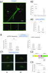
ApNT in the presynaptic neuron is important during the induction of ITF produced by 5HT. (A) ApNT is localized in both presynaptic and postsynaptic neurons of the sensorimotor neuron cocultures. (A1) Representative confocal microscope images of immunoreactivity (IR) using an antibody (M50) raised against mApNT compared with no antibody or the primary or secondary antibody alone. (Scale bar, 100 μm.) In this and subsequent figures, the SN cell body is ∼30 μm, and the MN cell body is ∼100 μm. (A2) There was a significant main effect of antibody on the intensity of ApNT immunostaing in a two-way ANOVA with one repeated measure, F(3, 8) = 11.29, P < 0.01. In addition, staining intensity was greater in optical sections of the presynaptic neuron than the postsynaptic neuron in the same dish (n = 6) (P < 0.05). (A3) Injection of siRNA against mApNT into the presynaptic neuron reduced the intensity of immunostaining for ApNT in the presynaptic SN (n = 5), compared with a control RNA injection (n = 5) (P < 0.01). (B) ApNT in the presynaptic neuron is involved in the increase in the frequency of mEPSCs during the induction of ITF by 5HT. Injection of siRNA against mApNT into the presynaptic neuron (n = 10) reduced the increase in mEPSC frequency produced by 5HT, compared with a control RNA injection (n = 11) (P < 0.001) without a clear effect on mEPSC amplitude (P > 0.05). The average pretest mEPSC values were 15 min−1 for frequency and 9 pA for amplitude, not significantly different between siRNA injection and control RNA injection groups. (C) 5HT stimulates the release of ApNT from SNs. (C1) Representative images before and 5 min after application of either 5HT (20 μM) or control solution to cocultures with SNs expressing ApNTpHluorin. (C2) Schematic diagram of the ApNTpHluorin construct. See the Results. (C3) Five-minute bath application of 5HT (n = 7) significantly increased the release of ApNTpHluorin from the presynaptic SN, compared with control (n = 9) (P < 0.05). *P < 0.05, **P < 0.01 for the difference between the experimental and control groups.
Fig. 3.
Fig. 3.
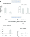
ApTrk in the presynaptic neuron is important during the induction of ITF produced by 5HT. (A) Injection of an antisense oligo against ApTrk into the SN (n = 7) reduced the increase in mEPSC frequency produced by 10 min 5HT, compared with sense oligo injection control (n = 4) (P < 0.05) without a clear effect on mEPSC amplitude (P > 0.05). The average pretest eEPSC values were 24 min−1 for frequency and 7 pA for amplitude, not significantly different between antisense oligo injection and sense oligo injection groups. (B) Activation of ApTrk receptor signaling in the presynaptic neuron increased mEPSC frequency. Shown are examples of mEPSCs recorded before and during the activation of presynaptic ApTrk signaling by application of BB dimers to cultures with SNs expressing ApTrkmem (SI Appendix, Figs. S1 and S2). There was a significant increase in mEPSC frequency in the BB ligand group (n = 9), compared with the control ligand group (n = 11) (P < 0.001) without a clear effect on mEPSC amplitude (P > 0.05). The average pretest values for mEPSC frequency were 8.5 min−1 for the BB group and 18.9 min−1 for the control group; the average mEPSC amplitude was 9.5 pA, not significantly different between the two groups. *P < 0.05, **P < 0.01 for the difference between the experimental and control groups.
Fig. 4.
Fig. 4.
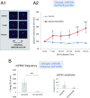
ApNT and ApTrk are downstream of PKA in the SN during the induction of ITF. (A1 and A2) Activation of OAR signaling coupled to PKA in the presynaptic neuron produced release of ApNT from the SN. (A1) Representative images before and 5 and 45 min after application of either OA (20 μM) or control solution to cocultures with SNs expressing OAR. (A2) Bath application of OA (n = 15) significantly increased the release of ApNTpHluorin from the presynaptic SN, compared with application of control solution (n = 15) (P < 0.05). The ApNTpHluorin signal gradually increased over time. (B) The increase in the frequency of mEPSCs produced by activation of OAR signaling coupled to PKA in the presynaptic neuron was reduced by injection of an antisense oligo against ApTrk receptors into the SN (n = 7), compared with sense oligo injection (n = 7) (P < 0.01) without a clear effect on mEPSC amplitude (P > 0.05). The average pretest mEPSC values were 24 min−1 for frequency and 7 pA for amplitude, not significantly different between the antisense oligo injection and sense oligo injection groups. xP < 0.05 one-tail, *P < 0.05, **P < 0.01 for the difference between the experimental and control groups.
Fig. 5.
Fig. 5.
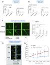
PKA, PKC, and Ca2+ are downstream of ApNT and ApTrk in the SN during the induction of ITF. (A1) Presynaptic PKA is involved in the signal transduction produced by mApNT. The increase in the frequency of mEPSCs produced by mApNT was significantly reduced by injection of a peptide inhibitor of PKA (PKAi 6–22) into the SN (n = 10), compared with vehicle injection (n = 13) (P < 0.001), without a clear effect on mEPSC amplitude (P > 0.05). The average pretest mEPSC values were 27.5 min−1 for frequency and 6 pA for amplitude, not significantly different between PKAi injection and vehicle injection. (A2) PKC in the presynaptic neuron is also involved in mApNT signaling. The increase in the frequency of mEPSCs produced by mApNT was significantly reduced by injection of a peptide inhibitor of PKC (PKCi 19–31) into the SN (n = 3), compared with vehicle injection (n = 13) (P < 0.05), without a clear effect on mEPSC amplitude (P > 0.05). The average pretest mEPSC values were 19.7 min−1 for frequency and 6.43 pA for amplitude, not significantly different between PKCi injection and vehicle injection. (B) Activation of presynaptic ApTrk receptors activates PKA in the presynaptic neuron. Application of BB dimers for 5 min to cocultures with SNs expressing ApTrk-mem (n = 10) increased immunostaining in the SNs with an antibody raised against the catalytic domain (active form) of PKA, compared with control treatment (n = 37) (P < 0.05). As positive controls, we also activated PKA in the presynaptic neuron either by stimulation of ectopically expressed OAR receptors in the presynaptic neuron (n = 7) or by bath application of 5HT (n = 19). Both manipulations also increased staining for the active form of PKA in the presynaptic neuron (P < 0.05 in each case). There was a significant main effect of group in a one-way ANOVA, F(3, 69) = 4.32, P < 0.01. (C1 and C2) Activation of presynaptic ApTrk receptors produced an increase in Ca2+ in the SN. (C1) Representative images of Calcium Orange fluorescence before and 5 and 45 min after application of either BB dimers or control solution to a coculture with a SN expressing ApTrk-mem. (C2) Bath application of BB dimers (n = 5) significantly increased Calcium Orange fluorescence in the presynaptic SN, compared with control (n = 10) (P < 0.05). The Ca2+ signal gradually increased over time. xP < 0.05 one-tail, *P < 0.05, ***P < 0.001 for the difference between the experimental and control groups.
Fig. 6.
Fig. 6.
Protein synthesis is downstream of ApNT and ApTrk in the SN during the induction of ITF. (A) Protein synthesis is required for the induction of ITF by mApNT. Bath application of anisomycin (n = 4) significantly reduced the increase in the frequency of mEPSCs produced by mApNT, compared with control (n = 9) (P < 0.05), without a clear effect on mEPSC amplitude (P > 0.05). The average pretest mEPSC values were 11.9 min−1 for frequency and 9.6 pA for amplitude, not significantly different between the anisomycin and control groups. (B) Protein synthesis is also required for the induction of ITF by activation of presynaptic ApTrk receptors. Bath application of anisomycin (n = 8) significantly reduced the increase in the frequency of mEPSCs produced by application of BB dimmers to cocultures with SNs expressing ApTrk-mem, compared with control (n = 4) (P < 0.05), without a clear effect on mEPSC amplitude (P > 0.05). The average pretest mEPSC values were 10.6 min−1 for frequency and 7.2 pA for amplitude, not significantly different between the anisomycin and control groups. (C1–C4) Activation of ApTrk receptors on the presynaptic neuron activates MAPK, AKT, and elF4E signaling pathways in the presynaptic neuron. (C1) Bath application of BB ligands to cocultures with SNs expressing ApTrk-mem (n = 4) significantly increased immunostaining for phospho-MAPK (p-MAPK) in the presynaptic SN, compared with control ligand (n = 9) (P < 0.01). BB dimer application (n = 4) also significantly increased immunostaining for phospho-AKT (p-AKT) in the presynaptic neuron, compared with control (n = 7) (P < 0.01). (C2) Activation of ApTrk receptors in the presynaptic neuron increased immunostaining for phospho-eIF4E (p-eIF4E), a major translational factor, in the presynaptic neuron. Bath application of BB dimers to cocultures with SNs expressing ApTrk-mem (n = 13) significantly increased immunostaining for p-eIF4E in the presynaptic SN, compared with control (n = 12) (P < 0.01). (C3 and C4) The immunostaining for both p-MAPK and p-eIF4E gradually increased over time.
Fig. 7.
Fig. 7.
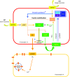
Presynaptic signaling pathways involved in the transition from STF to ITF. The induction of ITF involves presynaptic autocrine signaling by ApNT, which forms a presynaptic positive feedback loop with ApTrk receptors and PKA (pre-PKA → pre-ApNT → pre-ApTrk → pre-PKA). This feedback loop enhances spontaneous release of glutamate, which activates mGluR5 receptors and recruits postsynaptic mechanisms, and also stimulates protein synthesis in the presynaptic neuron, both of which are necessary for the transition to ITF. *Protein synthesis is thought to replenish the reserve vesicle pool and other synaptic proteins that support release of neurotransmitters from the presynaptic neuron.

Similar articles

Cited by

References

    1. Rosenzweig MR, Bennett EL, Colombo PJ, Lee DW, Serrano PA. Short-term, intermediate-term, and long-term memories. Behav Brain Res. 1993;57:193–198. - PubMed
    1. Raymond CR. LTP forms 1, 2 and 3: Different mechanisms for the “long” in long-term potentiation. Trends Neurosci. 2007;30:167–175. - PubMed
    1. Kandel ER, Dudai Y, Mayford MR. The molecular and systems biology of memory. Cell. 2014;157:163–186. - PubMed
    1. Hawkins RD, Mayford MR, Kandel ER. A comparative analysis of the molecular mechanisms contributing to implicit and explicit memory storage in Aplysia and in the hippocampus. In: Byrne JH, editor. Learning and Memory: A Comprehensive Reference. 2nd Ed. Elsevier; Oxford: 2016. pp. 5–32.
    1. Rayport SG, Schacher S. Synaptic plasticity in vitro: Cell culture of identified Aplysia neurons mediating short-term habituation and sensitization. J Neurosci. 1986;6:759–763. - PMC - PubMed

Publication types

MeSH terms

Substances