Review
. 2019 Feb;60(2):229-241.
doi: 10.1194/jlr.R089490. Epub 2018 Nov 5.

VPS34 complexes from a structural perspective

Affiliations
Review

VPS34 complexes from a structural perspective

Yohei Ohashi et al. J Lipid Res. 2019 Feb.

Abstract

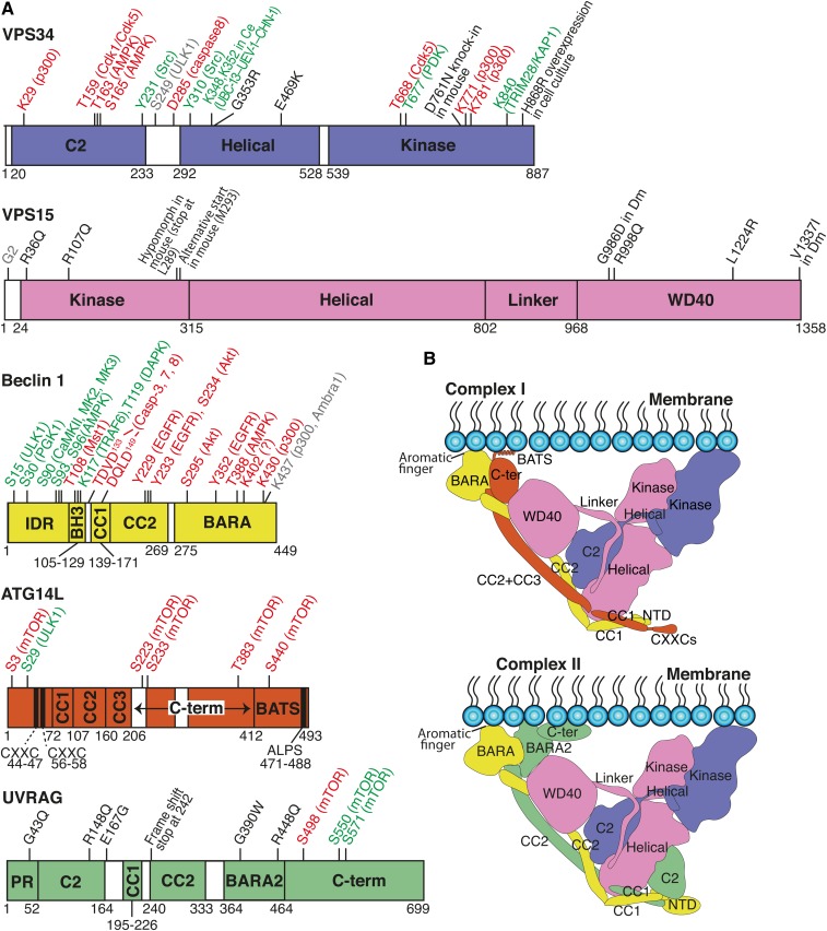
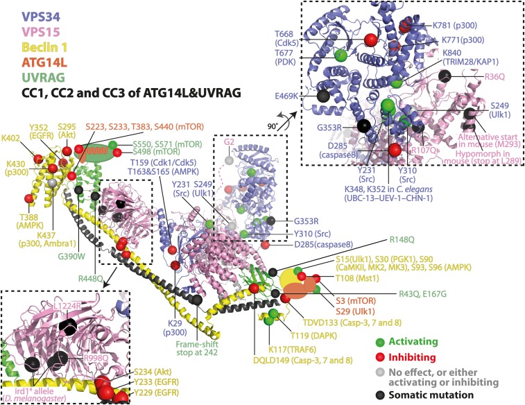
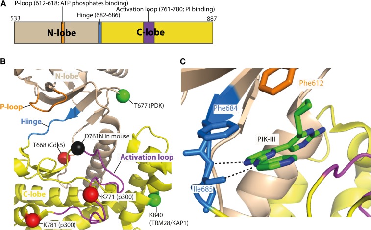
VPS34 phosphorylates phosphatidylinositol to produce PtdIns3P and is the progenitor of the phosphoinositide 3-kinase (PI3K) family. VPS34 has a simpler domain organization than class I PI3Ks, which belies the complexity of its quaternary organization, with the enzyme always functioning within larger assemblies. PtdIns3P recruits specific recognition modules that are common in protein-sorting pathways, such as autophagy and endocytic sorting. It is best characterized in two heterotetramers, complexes I and II. Complex I is composed of VPS34, VPS15, Beclin 1, and autophagy-related gene (ATG)14L, whereas complex II replaces ATG14L with UVRAG. Because VPS34 can form a component of several distinct complexes, it enables independent regulation of various pathways that are controlled by PtdIns3P. Complexes I and II are critical for early events in autophagy and endocytic sorting, respectively. Autophagy has a complex association with cancer. In early stages, it inhibits tumorigenesis, but in later stages, it acts as a survival factor for tumors. Recently, various disease-associated somatic mutations were found in genes encoding complex I and II subunits. Lipid kinase activities of the complexes are also influenced by posttranslational modifications (PTMs). Mapping PTMs and somatic mutations on three-dimensional models of the complexes suggests mechanisms for how these affect VPS34 activity.

Keywords: X-ray crystallography; cryo-electron microscopy; hydrogen-deuterium exchange mass-spectrometry; lipid; vacuolar protein sorting 34.


Figures

Fig. 1.
Fig. 1.
Structures of complex I and complex II. A: Schematic representations for the subunits of the class III PI3K complexes. Posttranslational modifcations (PTMs) and somatic mutations are indicated on the upper side of each subunit. Unless otherwise noted, all residue numbers are for the human sequences. Ce, C. elegans; Dm, D. melanogaster. Red, inhibiting; green, activating; gray, no effect or either inhibiting or activating; black, somatic mutations. B: Schematic structural models of complex I (top) and complex II (bottom). Because structural information on the CXXC, C-ter, and BATS regions of ATG14L, and the NTD and C-ter of UVRAG is not available, the boundaries of these domains are speculative.
Fig. 2.
Fig. 2.
Overall views of human complexes I and II. PTMs and somatic mutations are mapped on the yeast complex II structure [Protein Data Bank (PDB) identification 5DFZ) because this is the highest resolution and most complete structure. Human numbering is used unless otherwise noted. Dark gray, CC1+CC2 in UVRAG and CC1+CC2+CC3 in ATG14L.
Fig. 3.
Fig. 3.
Close-up views of the kinase domain in human VPS34. A: A schematic representation of the kinase domain in human VPS34. B: A structural view of the ATP-binding pocket of human VPS34 (PDB identification 3IHY). PTMs are indicated in red for inhibiting and green for activating, respectively. A mouse knock-in mutation (D761N) is indicated in black. C: An example of VPS34-specific inhibitor, PIK-III, binding to the hinge in the ATP-binding pocket (PDB identification 4PH4).

References

    1. Miller S., Tavshanjian B., Oleksy A., Perisic O., Houseman B., Shokat K., and Williams R.. 2010. Shaping development of autophagy inhibitors with the structure of the lipid kinase Vps34. Science. 327: 1638–1642. - PMC - PubMed
    1. Rostislavleva K., Soler N., Ohashi Y., Zhang L., Pardon E., Burke J. E., Masson G. R., Johnson C., Steyaert J., Ktistakis N. T., et al. . 2015. Structure and flexibility of the endosomal Vps34 complex reveals the basis of its function on membranes. Science. 350: aac7365. - PMC - PubMed
    1. Baskaran S., Carlson L-A., Stjepanovic G., Young L. N., Kim D. J., Grob P., Stanley R. E., Nogales E., and Hurley J. H.. 2014. Architecture and dynamics of the autophagic phosphatidylinositol 3-kinase complex. eLife. 3: e05115. - PMC - PubMed
    1. Stjepanovic G., Baskaran S., Lin M. G., and Hurley J. H.. 2017. Unveiling the role of VPS34 kinase domain dynamics in regulation of the autophagic PI3K complex. Mol. Cell. Oncol. 4: e1367873. - PMC - PubMed
    1. Stjepanovic G., Baskaran S., Lin M. G., and Hurley J. H.. 2017. Vps34 kinase domain dynamics regulate the autophagic PI 3-kinase complex. Mol. Cell. 67: 528–534.e3. - PMC - PubMed

Publication types

MeSH terms

Substances

LinkOut - more resources