. 2018 Dec;3(12):1377-1384.
doi: 10.1038/s41564-018-0271-y. Epub 2018 Nov 5.

A Chlamydia effector combining deubiquitination and acetylation activities induces Golgi fragmentation

Affiliations

A Chlamydia effector combining deubiquitination and acetylation activities induces Golgi fragmentation

Jonathan N Pruneda et al. Nat Microbiol. 2018 Dec.

Abstract

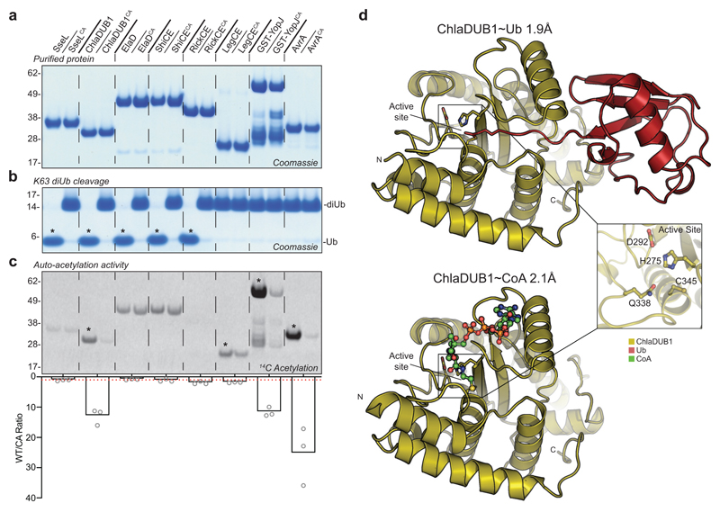
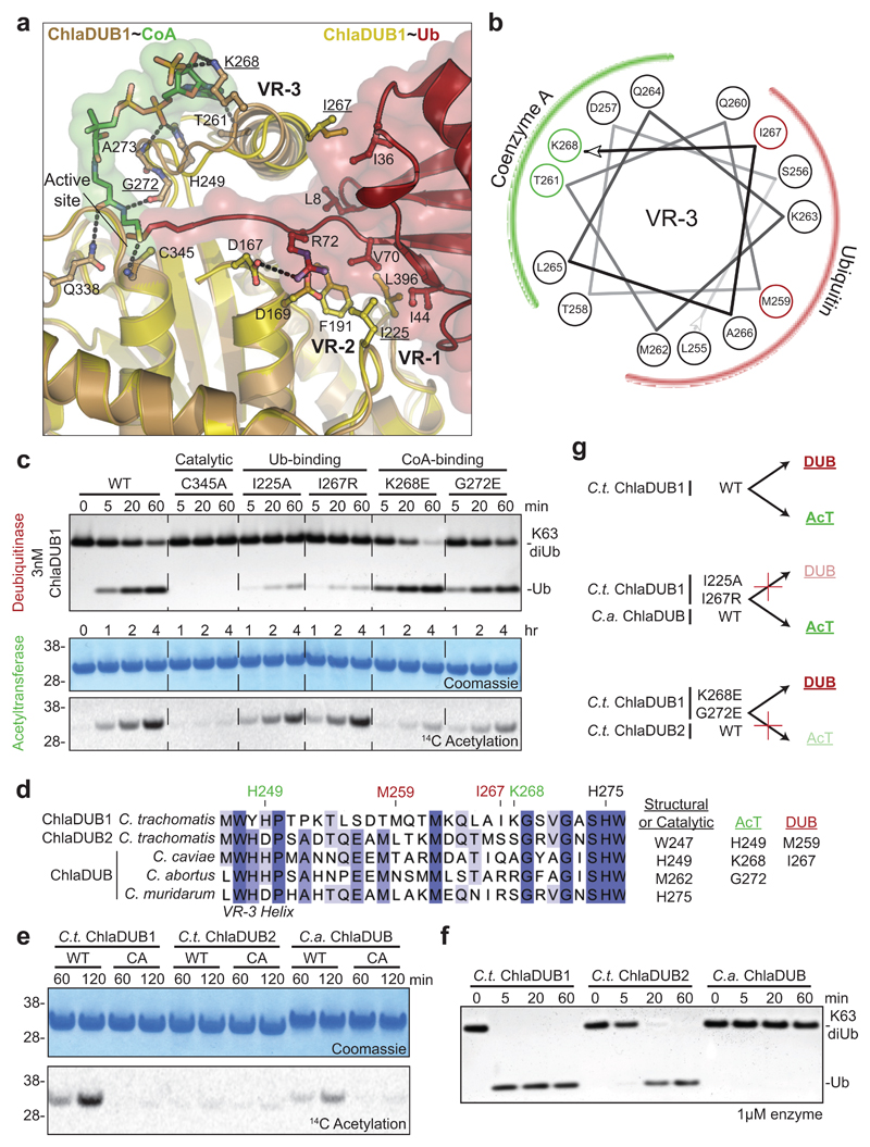
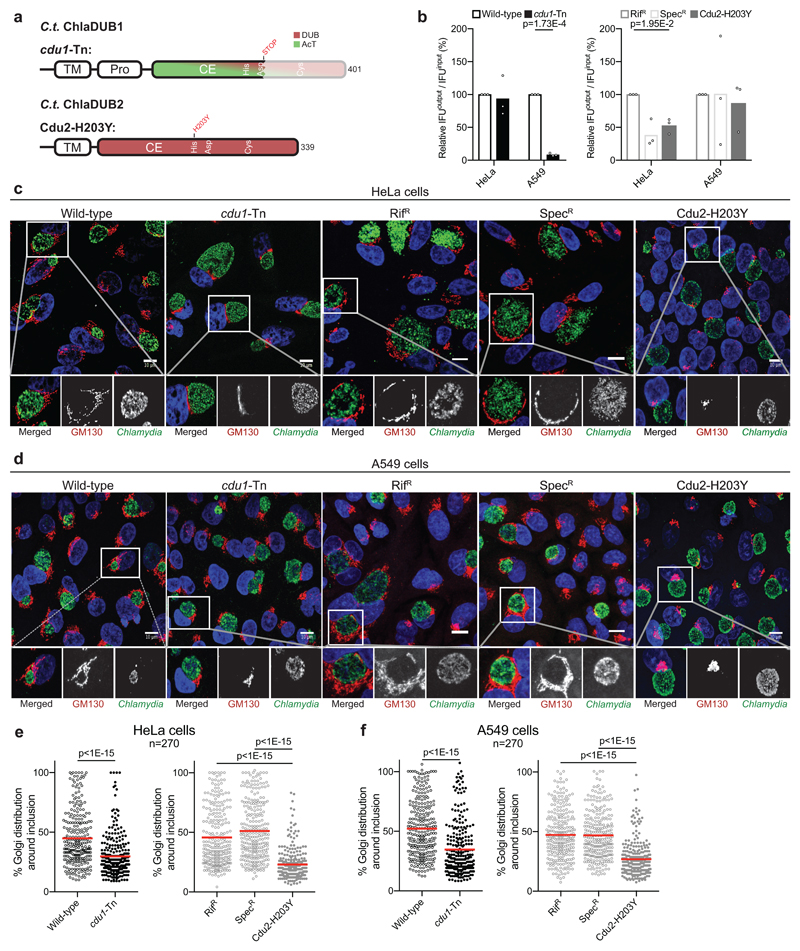
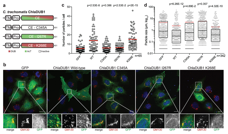
Pathogenic bacteria are armed with potent effector proteins that subvert host signalling processes during infection1. The activities of bacterial effectors and their associated roles within the host cell are often poorly understood, particularly for Chlamydia trachomatis2, a World Health Organization designated neglected disease pathogen. We identify and explain remarkable dual Lys63-deubiquitinase (DUB) and Lys-acetyltransferase activities in the Chlamydia effector ChlaDUB1. Crystal structures capturing intermediate stages of each reaction reveal how the same catalytic centre of ChlaDUB1 can facilitate such distinct processes, and enable the generation of mutations that uncouple the two activities. Targeted Chlamydia mutant strains allow us to link the DUB activity of ChlaDUB1 and the related, dedicated DUB ChlaDUB2 to fragmentation of the host Golgi apparatus, a key process in Chlamydia infection for which effectors have remained elusive. Our work illustrates the incredible versatility of bacterial effector proteins, and provides important insights towards understanding Chlamydia pathogenesis.


Conflict of interest statement

Competing Interests statement

The authors declare no competing interests.

Figures

Figure 1
Figure 1. Identification of specialized and dual-function CE-clan enzymes
a) Panel of purified bacterial CE-clan enzymes and their catalytically inactive Cys-to-Ala mutants. b) Deubiquitinase assay monitoring cleavage of K63-linked diUb following overnight incubation. c) Acetyltransferase assay monitoring 14C incorporation following a 2 h incubation of each protein with 14C-labeled Acetyl-CoA. Below, histogram representation of the WT/CA 14C incorporation ratio following normalization of the 14C autoradiography signal to the Coomassie stain. The average of three replicate experiments is plotted. A WT/CA ratio of one indicates no AcT activity, and is denoted by a red dashed line. Gels in a, b, and c are representative of triplicate experiments. All uncropped gels are shown in Supplementary Fig. 10. Asterisks indicate appreciable DUB (b) or AcT (c) activity. d) ChlaDUB1 complex crystal structures that capture intermediate stages of deubiquitinase (top) and acetyltransferase (bottom) activities. Inlay, a representative view of the ChlaDUB1 active site showing the Cys-His-Asp catalytic triad and the Gln oxyanion hole.
Figure 2
Figure 2. Molecular dissection of dual deubiquitinase/acetyltransferase activities
a) Close-up of the ChlaDUB1:CoA (brown:green) and ChlaDUB1:Ub (tan:red) interfaces with key interacting residues shown in ball-and-stick. Hydrogen bonds are shown as dashed lines. b) Helical wheel diagram illustrating the amphipathic nature of the ChlaDUB1 VR-3 helix, and its interactions with Ub or CoA (colored in red and green, respectively). c) Deubiquitinase (top) and acetyltransferase (bottom) assays illustrating that while both activities require the catalytic Cys residue, mutations in the Ub-binding and CoA-binding regions separate the two functions. A representative gel is shown of triplicate experiments. d) Sequence alignment of the VR-3 helix for orthologous Chlamydia ChlaDUB enzymes. The catalytic His, CoA-binding (green) and Ub-binding (red) residues are marked, and additional contacts are listed. e) 14C acetylation assay with the ChlaDUB orthologues from C. trachomatis (C.t. ChlaDUB2) and C. abortus (C.a. ChlaDUB). f) Deubiquitinase assay monitoring K63-linked diUb cleavage by the Chlamydia ChlaDUB orthologues. Gels in e and f are representative of triplicate experiments. All uncropped gels are shown in Supplementary Fig. 10. g) Schematic depicting how deubiquitinase and acetyltransferase functions can be separated either by structure-guided mutation or evolution as represented by the ChlaDUB orthologues.
Figure 3
Figure 3. ChlaDUB function is required for C. trachomatis Golgi fragmentation
a) Topology diagram illustrating C.t. ChlaDUB1 and C.t. ChlaDUB2 domain architecture, with active site residues annotated within the catalytic domains. Changes present in the cdu1-Tn and Cdu2-H203Y defective strains are shown above. b) C. trachomatis growth assay measured as inclusion forming units (IFU) output per IFU input following a 48 h infection in either HeLa or A549 cells. Values were normalized to 100% for wild-type. Statistical significance compared to parental controls was measured using a two-tailed Welch’s t-test. HeLa: Wild-type – cdu1-Tn, p=0.768; RifR – Cdu2-H203Y, p=0.0195; SpecR – Cdu2-H203Y, p=0.392. A549: Wild-type – cdu1-Tn, p=0.000173; RifR – Cdu2-H203Y, p=0.615; SpecR – Cdu2-H203Y, p=0.791. n=3. c) Representative confocal images showing Golgi fragmentation and redistribution around the Chlamydia inclusion following a 26 h infection of HeLa cells. Samples were immunostained with anti-GM130 (cis-Golgi, red) and anti-Slc1 (Chlamydia, green) antibodies, and Hoechst stained (DNA, blue). Isolated channels for the boxed region are shown below, and full versions are shown in Supplementary Fig. 6b. Scale bar corresponds to 10 µm. d) As in c) for A549 cells. Full versions are shown in Supplementary Fig. 6c. e) Quantification of c) following measurement of Golgi distribution around the circumference of the Chlamydia inclusion in 90 cells for each of three independent replicates. Mean values are shown as a red bar with individual data points overlaid. Statistical significance compared to parental was measured using a two-tailed Mann-Whitney test. Wild-type – cdu1-Tn, p<1E-15; RifR – Cdu2-H203Y, p<1E-15; SpecR – Cdu2-H203Y, p<1E-15. Separated plots for each replicate are shown in Supplementary Fig. 6d. f) As in e) for A549 cells. Wild-type –cdu1-Tn, p<1E-15; RifR – Cdu2-H203Y, p<1E-15; SpecR – Cdu2-H203Y, p<1E-15. Separated plots for each replicate are shown in Supplementary Fig. 6e.
Figure 4
Figure 4. ChlaDUB deubiquitinase activity is required for C. trachomatis Golgi fragmentation
a) Topology diagram illustrating the constructs used to characterize activity dependence of Golgi fragmentation following expression of ChlaDUB1 in mammalian cells. Separation-of-function mutations were selected from structural and biochemical work discussed in Fig. 2. b) Representative confocal images showing Golgi fragmentation in HeLa cells following expression of GFP-tagged ChlaDUB1. Samples were immunostained with anti-GM130 (cis-Golgi, red) and DAPI stained (DNA, blue). GFP fluorescence is shown in green. Isolated channels for the boxed region are shown below, and full versions are shown in Supplementary Fig. 7b. Scale bar corresponds to 10 µm. c) Quantification of cis-Golgi-stained puncta from b) for ~65 cells in each of three independent replicates (two remaining replicates are plotted in Supplementary Fig. 7c). Mean values are shown as red bars with individual data points overlaid. Statistical significance compared to GFP control was measured using a two-tailed Mann-Whitney test. GFP – WT, p=2.53E-8; GFP – C345A, p=0.386; GFP – I267R, p=0.0253; GFP – K268E, p=2E-15. d) Measurement of cis-Golgi-stained puncta size from b) for ~65 cells in each of three independent replicates (two remaining replicates are plotted in Supplementary Fig. 7d). Mean values are shown as red bars, median values are shown as black bars inside a quartile box plot, with individual data points overlaid. Statistical significance compared to GFP control was measured using a two-tailed Mann-Whitney test. GFP – WT, p=6.26E-12; GFP – C345A, p=0.0489; GFP – I267R, p=0.357; GFP – K268E, p=4.32E-10.

Similar articles

Cited by

References

    1. Lin YH, Machner MP. Exploitation of the host cell ubiquitin machinery by microbial effector proteins. J Cell Sci. 2017;130:1985–1996. - PMC - PubMed
    1. Bastidas RJ, Valdivia RH. Emancipating Chlamydia: Advances in the genetic manipulation of a recalcitrant pathogen. Microbiol Mol Biol Rev. 2016;80:411–427. - PMC - PubMed
    1. Rytkönen A, et al. SseL, a Salmonella deubiquitinase required for macrophage killing and virulence. Proc Natl Acad Sci USA. 2007;104:3502–3507. - PMC - PubMed
    1. Misaghi S, et al. Chlamydia trachomatis-derived deubiquitinating enzymes in mammalian cells during infection. Mol Microbiol. 2006;61:142–150. - PubMed
    1. Catic A, Misaghi S, Korbel GA, Ploegh HL. ElaD, a deubiquitinating protease expressed by E. coli. PLoS One. 2007;2:e381. - PMC - PubMed

Publication types

MeSH terms