. 2019 Jan;4(1):124-133.
doi: 10.1038/s41564-018-0275-7. Epub 2018 Nov 5.

Atomic structures of enterovirus D68 in complex with two monoclonal antibodies define distinct mechanisms of viral neutralization

Affiliations

Atomic structures of enterovirus D68 in complex with two monoclonal antibodies define distinct mechanisms of viral neutralization

Qingbing Zheng et al. Nat Microbiol. 2019 Jan.

Abstract

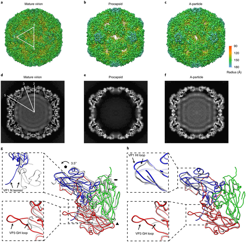
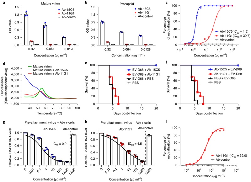
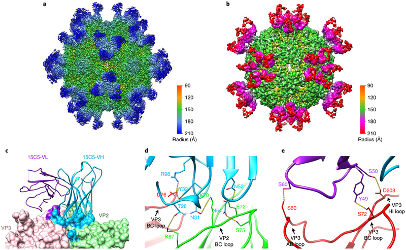
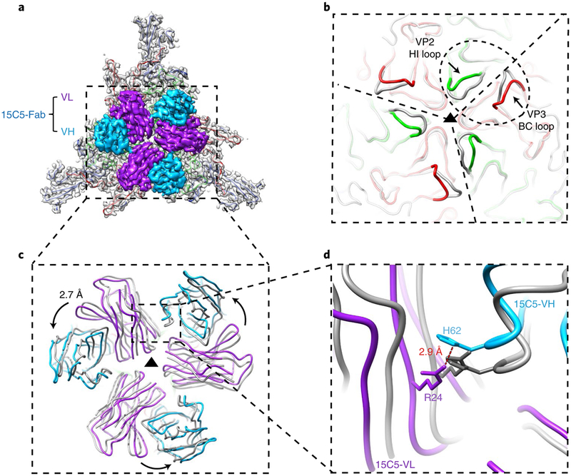
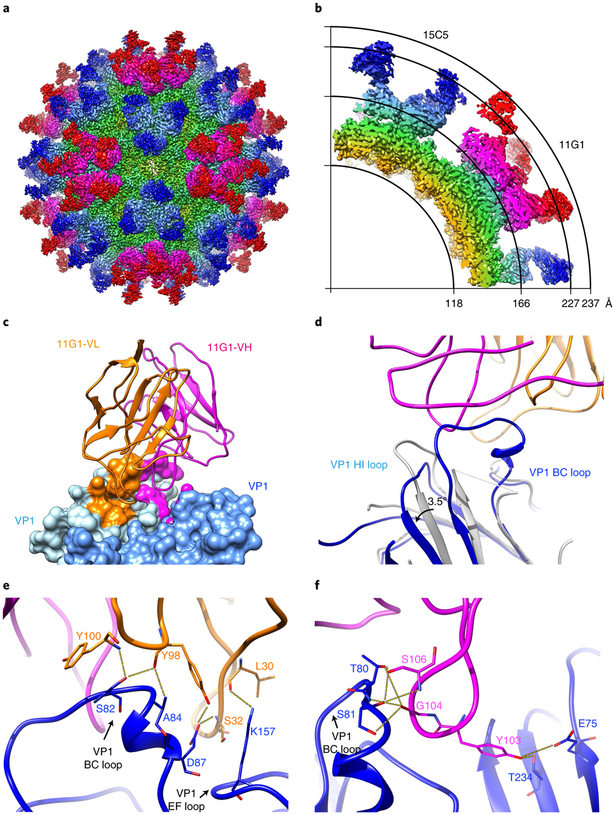
Enterovirus D68 (EV-D68) undergoes structural transformation between mature, cell-entry intermediate (A-particle) and empty forms throughout its life cycle. Structural information for the various forms and antibody-bound capsids will facilitate the development of effective vaccines and therapeutics against EV-D68 infection, which causes childhood respiratory and paralytic diseases worldwide. Here, we report the structures of three EV-D68 capsid states representing the virus at major phases. We further describe two original monoclonal antibodies (15C5 and 11G1) with distinct structurally defined mechanisms for virus neutralization. 15C5 and 11G1 engage the capsid loci at icosahedral three-fold and five-fold axes, respectively. To block viral attachment, 15C5 binds three forms of capsids, and triggers mature virions to transform into A-particles, mimicking engagement by the functional receptor ICAM-5, whereas 11G1 exclusively recognizes the A-particle. Our data provide a structural and molecular explanation for the transition of picornavirus capsid conformations and demonstrate distinct mechanisms for antibody-mediated neutralization.


Conflict of interest statement

Competing interests

The authors declare no competing interests.

Figures

Fig. 1 |
Fig. 1 |. The cryoEM structures of EV-D68 particles.
a–c, Iso-contoured views (along the icosahedral two-fold axis) of cryoEM maps (radially coloured) of the mature virion (a), procapsid (b) and A-particle (c). One icosahedral asymmetric unit is marked by a white triangle in a. The procapsid and A-particle show capsids with open two-fold channels, which are closed in the mature virion. d–f, Central sections of the corresponding maps displayed in the upper row. The densities of encapsidated genomic RNA are present in both the mature virion (d) and A-particle (f), but absent in the procapsid (e). g,h, Superpositions of protomers of A-particle protomer (VP1: blue; VP2: green; VP3: red) with that of mature virion (grey) (g) or procapsid (grey) (h). The orientation of the protomers is marked by black polygons indicating icosahedral five-, three- and two-fold axes, respectively. The colour scheme for the capsid proteins will be kept the same in all figures unless noted otherwise. The regions of the most significant differences between models are highlighted with magnified views in the dashed boxes: VP1 N termini (upper box) and VP3 GH loops (lower box) between the A-particle and the mature virion (g); VP1 HI loops (upper box) and VP3 GH loops (lower box) between the A-particle and the procapsid (h).
Fig. 2 |
Fig. 2 |. Characterization of the neutralizing antibodies 15C5 and 11G1.
a,b, Binding efficiencies of the neutralizing antibodies 15C5 and 11G1 to EV-D68 mature virion (a) or procapsid (b) evaluated with binding ELISA. The values are expressed as mean ± s.d. The experiments were independently repeated in triplicate, c, Neutralization efficiencies of the neutralizing antibodies 15C5 and 11G1 against EV-D68 virus plotted as a function of their concentrations (IC50 titres are noted), d, Particle stability thermal release assay of EV-D68 mature virions and their complexes with either 15C5 or 11G1. The fluorescence value indicates the level of genome exposure relative to temperature. The fluorescence traces are shown for EV-D68 mature virions (green line) as well as their complexes with 15C5 (blue line) or 11G1 (red line). The experiments were independently repeated in triplicate. e,f, Antibody-mediated protection against EV-D68 infections. Groups of 1-day-old BALB/c mice (10 mice per group) were treated with either 15C5 or 11G1 24 h after (e) or 12 h before (f) infection with EV-D68 (106 TCID50 per mouse, i.p.). The mortality of each group was monitored and recorded daily after infection. g,h, The amount of cell-bound EV-D68 viruses detected by quantitative real-time RT-PCR in the presence of the neutralizing antibody 15C5 (g) or 11G1 (h) added before the viruses. The values are expressed as mean ± s.d. The experiments were independently repeated in triplicate, i, Post-attachment neutralization assay of theneutralizing antibody 11G1 shows comparable neutralization capacity to that of regular conditions. The values are expressed as mean ± s.d. The experiments were repeated in triplicate. The IC50 was calculated by nonlinear regression fitting curves using GraphPad Prism version 7.0.
Fig. 3 |
Fig. 3 |. CryoEM structures of immune complexes EV-D68-M:15C5 and EV-D68-A:11G1.
a,b, Iso-contoured views of cryoEM maps (radially coloured) of the immune complexes EV-D68-M:15C5 (a) and EV-D68-A:11G1 (b). Groups of three 15C5 Fabs and five 11G1 Fabs bind at each three-fold and five-fold axis, respectively, c, Surface representation shows the interaction interface between Fab 15C5 and the capsid. Each Fab 15C5 binds across VP2 and VP3 from two adjacent protomers. Footprints of the VH and VL of the Fab on the capsid are coloured with light blue and purple, respectively. d,e, Close-up views of the interaction interface between the capsid and either the heavy (d) or light chain (e) of Fab 15C5. Note that the heavy chain binds across the VP2 BC loop and the VP3 BC loop from two adjacent protomers as shown in d. Potential hydrogen bonds and salt bridges are marked by yellow dashed lines.
Fig. 4 |
Fig. 4 |. Superposition of mature virion with or without the binding of 15C5 shows details of 15C5-induced conformational changes.
a, Outside view of the density attributable to the 3 three-fold-related asymmetric units segmented out of the cryoEM map of EV-D68-M:15C5. The capsid density (grey) is fitted with atomic models of three major capsid proteins and, for clarity, only the variable domains of three Fabs are shown, b, The 3 three-fold-related asymmetric units of EV-D68-M:15C5 (coloured wire diagram) are individually superimposed onto the models of the mature virion (grey). The scope of the epitope region is marked by the dashed ellipse. The significant conformational changes occurring at the VP2 HI loop and the VP3 BC loop after the binding of Fabs are highlighted. c,d, Atomic models of the three Fabs in a (coloured wire diagram) together with three superimposed Fabs (grey). The superimposed Fabs, which show a 2.7 Å movement and became closer when compared to the original model of the 15C5 immune complex, represent the theoretical binding of Fabs to the mature virion at the same epitope prior to conformational changes of the capsid. The close-up view (d) shows that the two nearest residues (distance of 2.9 Å) between neighbouring heavy and light chains are His 62H and Arg 24L derived from two adjacent Fabs. These two amino acids exhibit a steric clash in the superimposed models (grey).
Fig. 5 |
Fig. 5 |. The cryoEM structure of EV-D68 in complex with 15C5 and 11G1.
a, Iso-contoured views of the cryoEM map (radially coloured) of the immune complex EV-D68-M:15C5:11G1. The EV-D68 capsid is bound with 60 copies of Fab 15C5 at three-fold axes and 60 copies of Fab 11G1 at five-fold axes, respectively, b, A quarter of the central section of the cryoEM map in a; the radii are marked by the arcs, c, Surface representation shows the interaction interface of Fab 11G1. Each Fab 11G1 (heavy chain: magenta; light chain: orange) binds across two neighbouring VP1s (light blue and blue) of the capsid. Footprints of the VH and VL of the Fab on the capsid are coloured with magenta and orange, d, A close-up view (wire diagram) of the interaction interface between 11G1 and the capsid shows the conformational changes (indicated with black arrows) of the VP1 BC loop and HI loop before (grey) and after (coloured) binding with 11G1. e,f, Close-up views of the interaction interfaces between the capsid and the light (e) or heavy chain (f) of Fab 11G1. Potential hydrogen bonds are marked by yellow dashed lines.

Similar articles

Cited by

References

    1. Schieble JH, Fox VL & Lennette EH A probable new human picornavirus associated with respiratory diseases. Am. J. Epidemiol 85, 297–310 (1967). - PubMed
    1. Ikeda T et al. Acute respiratory infections due to enterovirus 68 in Yamagata, Japan between 2005 and 2010. Microbiol. Immunol 56, 139–143 (2012). - PubMed
    1. Kaida A et al. Distinct genetic clades of enterovirus D68 detected in 2010, 2013, and 2015 in Osaka City, Japan. PLoS ONE 12, e0184335 (2017). - PMC - PubMed
    1. Knoester M et al. Upsurge of enterovirus D68, the Netherlands, 2016. Emerg. Infect. Dis 23, 140–143 (2017). - PMC - PubMed
    1. Midgley CM et al. Severe respiratory illness associated with enterovirus D68 - Missouri and Illinois, 2014. MMWR. Morb. Mortal. Wkly Rep. 63, 798–799 (2014). - PMC - PubMed

Publication types