

Efficacy of Caries Removal by Carie-Care and Erbium-doped Yttrium Aluminum Garnet Laser in Primary Molars: A Scanning Electron Microscope Study
- PMID: 30397377
- PMCID: PMC6212662
- DOI: 10.5005/jp-journals-10005-1533
Efficacy of Caries Removal by Carie-Care and Erbium-doped Yttrium Aluminum Garnet Laser in Primary Molars: A Scanning Electron Microscope Study
Abstract
Aim: To compare and evaluate morphological changes and bacterial deposits in primary carious molars after caries excavation with Carie-Care, erbium-doped yttrium aluminum garnet (Er:YAG) laser, and round tungsten carbide bur.
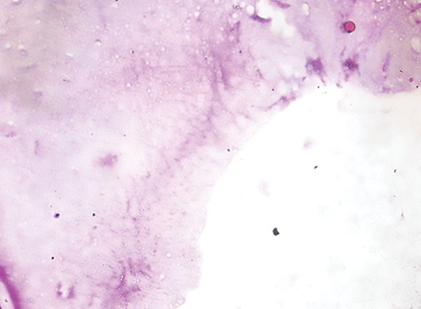
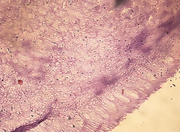
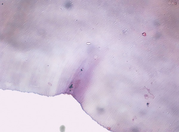
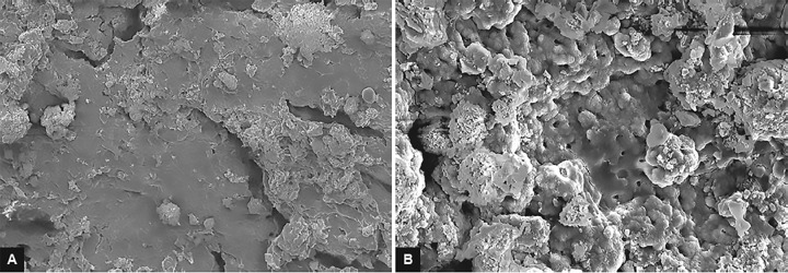
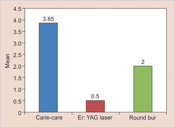
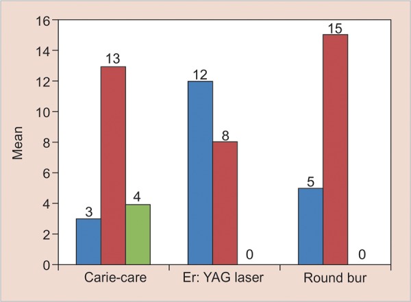
Materials and methods: Thirty human carious primary molars extracted for therapeutic reasons were sectioned mesiodistally. These sectioned samples were allocated into three groups (20 samples each): group I: Carie Care, group II: Er:YAG laser, and group III: round tungsten carbide bur. After caries excavation, all samples were processed and examined under conventional light microscope to examine for bacterial deposits. Representative samples from each group were processed and analyzed to examine the morphology of caries-excavated tissue by scanning electron microscope (SEM). Statistical analysis was done using Fisher's exact test, Kruskal-Wallis test, and Mann-Whitney U test.
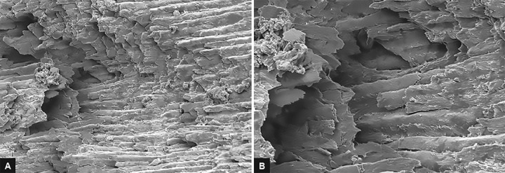
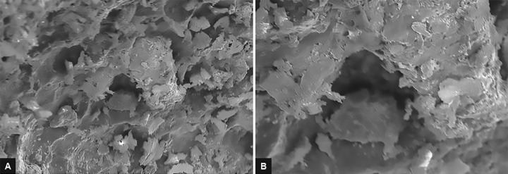
Results: The Er:YAG laser showed best results with no smear layer followed by chemomechanically excavated surfaces with Carie-Care. Amount of bacterial deposits was observed to be more in group I while least in group II (p-value < 0.001). Mann-Whitney U test and Fisher's exact test revealed that there was statistically significant difference among all the three groups.
Conclusion: Among the three different methods of caries excavation, Er:YAG laser was found to be more effective compared with Carie-Care and round tungsten carbide bur.
Clinical significance: Laser-induced caries excavation by Er:YAG laser and chemomechanical method of caries removal by Carie-Care can be considered as future of noninvasive pediatric and preventive dentistry.How to cite this article: Prabhakar A, Lokeshwari M, Naik SV, Yavagal C. Efficacy of Caries Removal by Carie-Care and Erbium-doped Yttrium Aluminum Garnet Laser in Primary Molars: A Scanning Electron Microscope Study. Int J Clin Pediatr Dent 2018;11(4):323-329.
Keywords: Carie-Care; Caries excavation; Carious primary molars; Erbium-doped yttrium aluminum garnet laser; Light microscopy; Round tungsten carbide bur; Scanning electron microscope..
Conflict of interest statement
Source of support: Nil Conflict of interest: None
Figures
Similar articles
-
Evaluation of the Erbium-doped Yttrium Aluminum Garnet Laser and the Conventional Method on Pain Perception and Anxiety Level in Children during Caries Removal: A Randomized Split-mouth Study.Int J Clin Pediatr Dent. 2023 Aug;16(Suppl 1):S39-S44. doi: 10.5005/jp-journals-10005-2634. Int J Clin Pediatr Dent. 2023. PMID: 37663205 Free PMC article.
-
Comparison of Marginal Microleakage of Glass Ionomer Restorations in Primary Molars Prepared by Chemo-mechanical Caries Removal (CMCR), Erbium: Yttrium Aluminum-Garnet (Er:YAG) Laser and Atraumatic Restorative Technique (ART).Int J Clin Pediatr Dent. 2013 May;6(2):75-9. doi: 10.5005/jp-journals-10005-1193. Epub 2013 Aug 26. Int J Clin Pediatr Dent. 2013. PMID: 25206196 Free PMC article.
-
Assessment of Smear Layer Formation After Caries Removal Using Erbium Laser and Papain-Based Chemo-Mechanical Caries Removal Agent: An In Vitro Scanning Electron Microscopy Study.Cureus. 2023 Oct 30;15(10):e47999. doi: 10.7759/cureus.47999. eCollection 2023 Oct. Cureus. 2023. PMID: 38034221 Free PMC article.
-
A Comparative Evaluation of the Efficacy of Different Caries Excavation Techniques in reducing the Cariogenic Flora: An in vivo Study.Int J Clin Pediatr Dent. 2016 Jul-Sep;9(3):214-217. doi: 10.5005/jp-journals-10005-1366. Epub 2016 Sep 27. Int J Clin Pediatr Dent. 2016. PMID: 27843252 Free PMC article. Review.
-
Efficacy and Patient's Acceptance of Alternative Methods for Caries Removal-a Systematic Review.J Clin Med. 2020 Oct 23;9(11):3407. doi: 10.3390/jcm9113407. J Clin Med. 2020. PMID: 33114249 Free PMC article. Review.
Cited by
-
Chemomechanical caries removal methods: A literature review.Saudi Dent J. 2023 Mar;35(3):233-243. doi: 10.1016/j.sdentj.2023.01.010. Epub 2023 Feb 6. Saudi Dent J. 2023. PMID: 37091279 Free PMC article. Review.
-
Assessment of the efficiency of dental excavation methods using laser speckle imaging.Lasers Med Sci. 2024 May 25;39(1):137. doi: 10.1007/s10103-024-04094-z. Lasers Med Sci. 2024. PMID: 38795227
-
The Use of Modern Technologies by Dentists in Poland: Questionnaire among Polish Dentists.Healthcare (Basel). 2022 Jan 25;10(2):225. doi: 10.3390/healthcare10020225. Healthcare (Basel). 2022. PMID: 35206840 Free PMC article.
-
An in vitro assessment of the residual dentin after using three minimally invasive caries removal techniques.Sci Rep. 2024 Mar 26;14(1):7087. doi: 10.1038/s41598-024-57745-0. Sci Rep. 2024. PMID: 38528204 Free PMC article.
-
The effectiveness of Carie-Care™, chemomechanical caries removal technique in primary teeth: randomized controlled clinical trial.BMC Oral Health. 2023 Nov 18;23(1):882. doi: 10.1186/s12903-023-03594-8. BMC Oral Health. 2023. PMID: 37980471 Free PMC article. Clinical Trial.
References
-
- Yip HK, Samaranayake LP. Caries removal techniques and instrumentation: a review. Clin Oral Investig. 1998 Dec;2(4):148–154. - PubMed
-
- Celiberti P, Francescut P, Lussi A. Performance of four dentine excavation methods in deciduous teeth. Caries Res. 2006;40(2):117–123. - PubMed
-
- Garcia-Contreras R, Scougall-Vilchis RJ, Contreras-Bulnes R, Sakagami H, Morales-Luckie RA, Nakajima H. A comparative in vitro efficacy of conventional rotary and chemomechanical caries removal: Influence on cariogenic flora, microhardness, and residual composition. J Conserv Dent. 2014 Nov;17(6):536–540. - PMC - PubMed
-
- Rajakumar S, Mungara J, Joseph E, Philip J, Guptha V, Shilpa Pally MP. Evaluation of three different caries removal techniques in children: a comparative clinical study. J Clin Pediatr Dent. 2013 Fall;38(1):23–26. - PubMed
LinkOut - more resources
Full Text Sources