. 2018 Oct 30:9:2041731418808695.
doi: 10.1177/2041731418808695. eCollection 2018 Jan-Dec.

Magnetic ion channel activation of TREK1 in human mesenchymal stem cells using nanoparticles promotes osteogenesis in surrounding cells

Affiliations

Magnetic ion channel activation of TREK1 in human mesenchymal stem cells using nanoparticles promotes osteogenesis in surrounding cells

James R Henstock et al. J Tissue Eng. .

Abstract

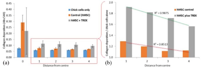
Magnetic ion channel activation technology uses superparamagnetic nanoparticles conjugated with targeting antibodies to apply mechanical force directly to stretch-activated ion channels on the cell surface, stimulating mechanotransduction and downstream processes. This technique has been reported to promote differentiation towards musculoskeletal cell types and enhance mineralisation. Previous studies have shown how mesenchymal stem cells injected into a pre-mineralised environment such as a foetal chick epiphysis, results in large-scale osteogenesis at the target site. However, the relative contributions of stem cells and surrounding host tissue has not been resolved, that is, are the mesenchymal stem cells solely responsible for the observed mineralisation or do mechanically stimulated mesenchymal stem cells also promote a host-tissue mineralisation response? To address this, we established a novel two-dimensional co-culture assay, which indicated that magnetic ion channel activation stimulation of human mesenchymal stem cells does not significantly promote migration but does enhance collagen deposition and mineralisation in the surrounding cells. We conclude that one of the important functions of injected human mesenchymal stem cells is to release biological factors (e.g., cytokines and microvesicles) which guide the surrounding tissue response, and that remote control of this signalling process using magnetic ion channel activation technology may be a useful way to both drive and regulate tissue regeneration and healing.

Keywords: Magnetic nanoparticles; mesenchymal stem cell; paracrine; stretch-activated ion channel; tissue engineering.


Conflict of interest statement

Declaration of conflicting interests: The author(s) declared no potential conflicts of interest with respect to the research, authorship and/or publication of this article.

Figures

Figure 1.
Figure 1.
MICA activation of the TREk1 stretch-activated ion channel. (a) Superparamagnetic ion oxide nanoparticles (SPIONS) were surface functionalised with antibodies specific to the mechanosensitive intracellular loop region of the TREK1 ion channel. (b) Attachment of the nanoparticle to the ion channel allows the ion channel to be activated (opened) using an external magnetic field. (c) Tagging TREK1 in hMSCs allows remote control of mechanotransduction using magnets, such as the (i) MICA bioreactor moving magnetic array used in this investigation, and (ii) remote control of injected hMSCs as reported by Henstock et al.
Figure 2.
Figure 2.
Magnetic field gradients. Graphical representation of the magnetic field gradients of the six-well permanent array used in this investigation. When the array was positioned towards the detector/culture, this resulted in a peak magnetic field range of 240–400 mT. Away from the sample, at a distance of 84 mm, the magnetic field strength was reduced to 1–2 mT. The images in red (inset) show representations of the field pattern experienced at the well base at each distance from the array.
Figure 3.
Figure 3.
Six-well array magnetic field mapping. 2D model representations of magnetic field strength and polarity of six-well permanent magnet arrays. When the magnetic array was closest to the samples (approximately 18 mm), the peak magnetic field strength was ±75 mT (a), the peak field strength and field polarity is shown in (c). A representative cross-section of the magnetic fields and polarity is shown in (e). When the magnetic array was the furthest (measurable) distance from the samples (approximately 84 mm), the magnetic field strength was reduced to ±1.4 mT (b), the field strength and field polarity is shown in (d). A representative cross-section of the magnetic fields and polarity is shown in (f).
Figure 4.
Figure 4.
Transwell migration of hMSCs tagged with magnetic nanoparticles. PKH26-labelled mesenchymal stem cells (hMSCs) (103 cells) were placed in the upper chamber of FluoroBlok transwell and migration across the membrane was quantified after 96 h. The relative migration of hMSCs labelled with either unconjugated (blank) or TREK1-targeting nanoparticles was compared to unlabelled cells (control) and unlabelled cells exposed to the magnet. High concentration foetal calf serum (30% FCS) served as the positive control. All groups exposed to the nanoparticles or the magnetic array showed a slight (non-significant) increase in migration over the control, and the TREK1-stimulated group (highlighted red) showed significantly increased migration (p = 0.02) over the control. Bars represent the experimental mean, and error bars show standard deviation, n = 6. *p < 0.05 and ***p < 0.001.
Figure 5.
Figure 5.
Migration of hMSCs into a co-culture of chick epiphyseal chondrocytes was not significantly enhanced by mechanotransduction. (a) PKH26-labelled hMSCs (103) were placed in the centre of a well in a six-well plate and 106 chick epiphyseal cells were seeded on top. The outgrowth of the PKH26-labelled hMSCs (b) from the central seeding dot was quantified after 28 days using the area scan function (c) of a fluorescence plate reader. (d) No significant migration was observed in either the control or TREK1 nanoparticle-tagged hMSCs. (e) Statistical analysis showed that there was no significant migration across the plate, and that the hMSCs remained in the same place for the duration of the experiment (28 days). (f) Error bars show standard deviation, n = 6.
Figure 6.
Figure 6.
Collagen deposition by chick epiphyseal cells was enhanced by TREK1 nanoparticle–activated hMSCs. (a) Collagen production was increased by ~20% when chick epiphyseal cells were cultured with hMSCs. When hMSCs are mechanically stimulated by TREK1 magnetic nanoparticles, collagen production (predominantly from the surrounding chick cells) was increased by 60%–90%. (b) A closer examination of collagen deposition as a function of distance from the central hMSC colony shows that chick epiphyseal cells closer to the hMSCs produced more collagen (normalised against the chick-only control and presented as fold-change). Collagen production by chick cells was inversely proportional to their distance from the hMSCs, indicating a biochemical concentration gradient or direct cell–cell contact is required to stimulate upregulation of collagen synthesis. Error bars show standard deviation, n = 6; R2 values indicate coefficient of linear regression.
Figure 7.
Figure 7.
Mineralisation in the co-culture was significantly increased by the presence of hMSCs and activation of hMSC mechanotransduction. (a) After 28 days, calcium deposition was determined by staining the plates with alizarin red, which was then solubilised with cetylpyridinium chloride and quantified spectrometrically. Calcium deposition was enhanced by the presence of the hMSCs (37% increase in total calcium deposition) and further enhanced by the activation of hMSC mechanotransduction (128% increase in calcium deposition) compared to the controls lacking MSCs. Visual analysis of the mineral distribution (b) shows that the chick epiphyseal cells mineralise the extracellular matrix (i) and mineralisation in the groups containing MSCs was localised in the central region surrounding the hMSCs (ii) and TREK-activated MSCs (iii) – the central zone delimiting the MSCs is indicated by the blue circles. At the 28 day end point, there was no significant difference in alkaline phosphatase activity in the cell-culture media supernatant (c). Error bars show standard deviation, n = 6 and ***p < 0.001.

References

    1. Henstock JR, Hu B, Markides H, et al. Applications of magnetic nanoparticles in tissue engineering and regenerative medicine. In: Dobson J. (ed.) Nanomagnetic actuation of cell surface receptors: basic principles and applications. Boca Raton: Taylor & Francis, 2018.
    1. Markides H, McLaren JS, Telling ND, et al. Translation of remote control regenerative technologies for bone repair. Regen Med 2018; 3: 9. - PMC - PubMed
    1. Henstock JR, El Haj AJ. Remotely controlled magnetic nanoparticles for bone regeneration. Regen Med 2015; 10: 377–380. - PubMed
    1. Henstock JR, Rotherham M, Rashidi H, et al. Remotely activated mechanotransduction via magnetic nanoparticles promotes mineralisation synergistically with BMP2: applications for injectable cell therapy. Stem Cell Transl Med 2014; 3: 1–12. - PMC - PubMed
    1. Hughes S, McBain S, Dobson J, et al. Selective activation of mechanosensitive ion channels using magnetic particles. J R Soc Interface 2008; 6: 855–863. - PMC - PubMed

LinkOut - more resources