

Giant solitary fibrous tumor of the pleura
- PMID: 30397437
- PMCID: PMC6210664
- DOI: 10.1093/jscr/rjy270
Giant solitary fibrous tumor of the pleura
Abstract
Introduction: Solitary fibrous tumors of the pleura (SFTP) are rare mesenchymal tumors representing <5% of all tumors of the pleura. Literature reveals only two case series and a few solitary reports.
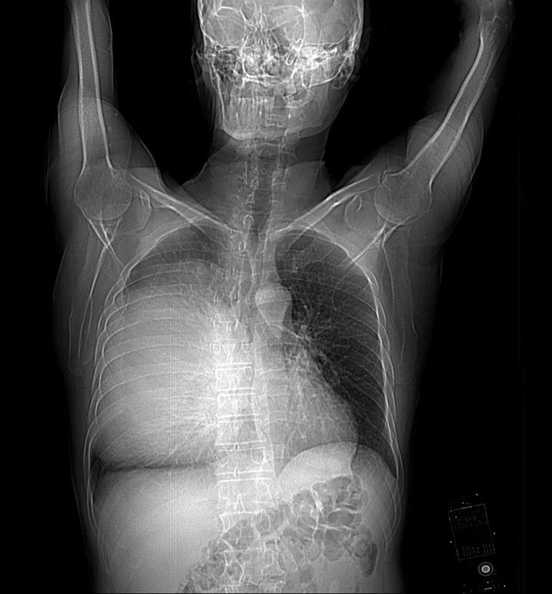
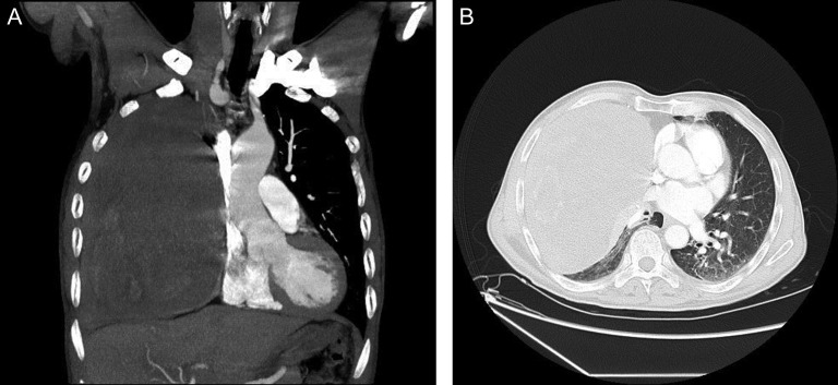
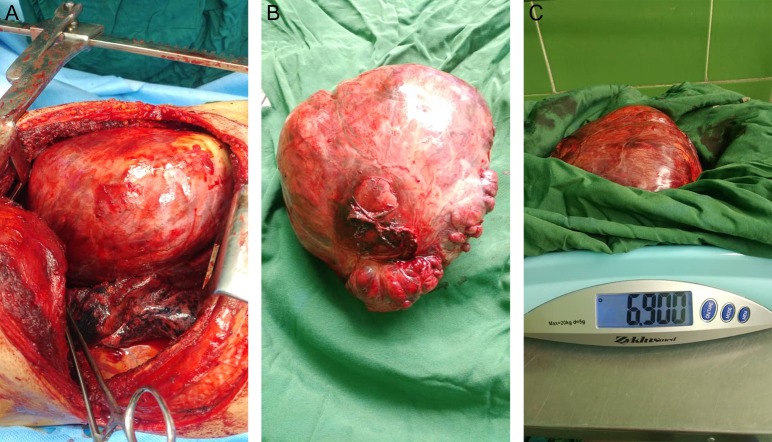
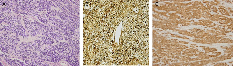
Case report: A 68-year-old male presented to our hospital after experiencing exertional dyspnea. A chest CT revealed a giant heterogeneous mass. CT-guided transthoracic core needle biopsy demonstrated SFTP. The well-circumscribed, encapsulated resected mass was measured to be 30 cm × 21 cm × 15 cm and weighed 6900 g.
Discussion: SFTP are a rare pathology of the pleural cavity, which most of the time develop from submesothelial fibroblasts of the visceral pleura. Due to their non-characteristic clinical picture, SFTP are usually diagnosed in the later stages of the development. A significant issue in the management of giant SFTP is radical resection of the tumor to relieve compression of the lung parenchyma and other mediastinal structures.
Conclusion: SFTP are rare neoplasms that fortunately are benign 80% of the time. Only a few cases of giant SFTP that cover almost the entire pleural space are described in the literature. This report represents one of the largest resected SFTP reports in the literature.
Figures
References
-
- Cardillo G, Facciolo F, Cavazzana AO, Capece G, Gasparri R, Martelli M. Localized(solitary) fibrous tumors of the pleura: an analysis of 55 patients. Ann Thorac Surg 2000;70:1808–12. - PubMed
-
- England DM, Hochholzer L, McCarthy MJ. Localized benign ena malignant fibrous tumor of pleura: a clinicopathologic review of 223 cases. Am J Pathol 1989;13:640–58. - PubMed
-
- Dynes MC, White EM, Fry WA, Ghahremany GG. Imaging manifestations of pleural tumors. Radiographics 1992;12:1191–201. - PubMed
Publication types
LinkOut - more resources
Full Text Sources
