

Live birth after hysteroscopy performed inadvertently during early pregnancy: A case report and review of literature
- PMID: 30397614
- PMCID: PMC6212613
- DOI: 10.12998/wjcc.v6.i12.559
Live birth after hysteroscopy performed inadvertently during early pregnancy: A case report and review of literature
Abstract
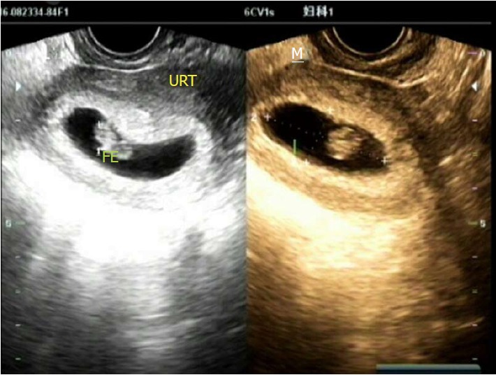
Generally, hysteroscopy is not appropriate for pregnant women without an indication. What if a patient undergoes hysteroscopy accidentally during the early gestational period? We here report a rare case of a woman who continued pregnancy after a diagnostic hysteroscopy was performed in early pregnancy and delivered a healthy baby. The patient had a history of infertility and oligomenorrhea, probably due to a previous induced abortion. A hysteroscopy was performed after the end of her "menstruation" for assessment of her uterine cavity. Early pregnancy, instead of the expected intrauterine adhesions, was suspected, and the procedure was immediately ceased. Subsequent tests confirmed the diagnosis of pregnancy. She had a full-term delivery by elective caesarean section. The success of this case was attributed to the use of vaginoscopic techniques in hysteroscopy and correct judgment and decision-making during the procedure. This case report provides some useful methods and experience that might be helpful when a similar situation occurs in clinical practice.
Keywords: Gestation; Hysteroscopy; Livebirth; Ongoing pregnancy.
Conflict of interest statement
Conflict-of-interest statement: The authors declare that they have no conflicts of interests to disclose.
Figures


Similar articles
-
Hysteroscopy as an aid to diagnosis in female infertility.Clin Obstet Gynecol. 1983 Jun;26(2):302-12. doi: 10.1097/00003081-198306000-00010. Clin Obstet Gynecol. 1983. PMID: 6851287
-
Efficiency and pregnancy outcome of serial intrauterine device-guided hysteroscopic adhesiolysis of intrauterine synechiae.Fertil Steril. 2008 Nov;90(5):1973-7. doi: 10.1016/j.fertnstert.2007.06.074. Epub 2008 Sep 6. Fertil Steril. 2008. PMID: 18774563 Clinical Trial.
-
Live birth rate and obstetric complications following the hysteroscopic management of intrauterine adhesions including Asherman syndrome.Hum Reprod. 2018 Oct 1;33(10):1847-1853. doi: 10.1093/humrep/dey237. Hum Reprod. 2018. PMID: 30239778
-
Fertility after contraception or abortion.Fertil Steril. 1990 Oct;54(4):559-73. doi: 10.1016/s0015-0282(16)53808-4. Fertil Steril. 1990. PMID: 2209874 Review.
-
Intrauterine adhesions.Obstet Gynecol Clin North Am. 1995 Sep;22(3):491-505. Obstet Gynecol Clin North Am. 1995. PMID: 8524533 Review.
References
-
- Salazar CA, Isaacson KB. Office Operative Hysteroscopy: An Update. J Minim Invasive Gynecol. 2018;25:199–208. - PubMed
-
- Assaf A, Gohar M, Saad S, el-Nashar A, Abdel Aziz A. Removal of intrauterine devices with missing tails during early pregnancy. Contraception. 1992;45:541–546. - PubMed
-
- Hooker A, Fraenk D, Brölmann H, Huirne J. Prevalence of intrauterine adhesions after termination of pregnancy: a systematic review. Eur J Contracept Reprod Health Care. 2016;21:329–335. - PubMed
-
- Taşkın EA, Berker B, Ozmen B, Sönmezer M, Atabekoğlu C. Comparison of hysterosalpingography and hysteroscopy in the evaluation of the uterine cavity in patients undergoing assisted reproductive techniques. Fertil Steril. 2011;96:349–352.e2. - PubMed
-
- akas P, Hassiakos D, Grigoriadis C, Vlahos N, Liapis A, Gregoriou O. Role of hysteroscopy prior to assisted reproduction techniques. J Minim Invasive Gynecol. 2014;21:233–237. - PubMed
Publication types
LinkOut - more resources
Full Text Sources
