

Mesh migration into the sigmoid colon after inguinal hernia repair presenting as a colonic polyp: A case report and review of literature
- PMID: 30397615
- PMCID: PMC6212604
- DOI: 10.12998/wjcc.v6.i12.564
Mesh migration into the sigmoid colon after inguinal hernia repair presenting as a colonic polyp: A case report and review of literature
Abstract
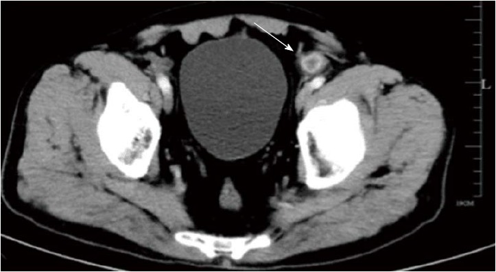
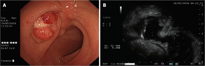
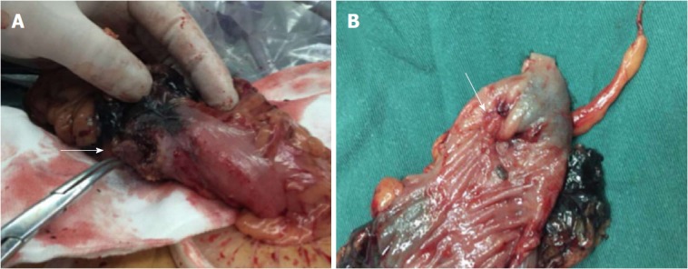
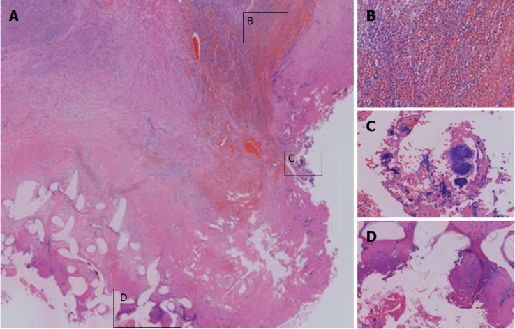
Mesh migration and penetration into abdominal viscera rarely occur after laparoscopic inguinal hernia repair. We present the first case of mesh migration into the sigmoid colon identified as a colonic polyp at initial colonoscopic examination. The patient complained of mild abdominal distention in the lower abdomen over the previous year without changes in bowel habits or stool appearance and without weight loss. By complementary endoscopic ultrasonography, a cavity-like structure beneath the suspected polyp was further confirmed. Enhanced abdominal computed tomography merely revealed local bowel wall thickening and inflammation of the colosigmoid junction. The migrating mesh, which was lodged in the sigmoid colon and caused intra-abdominal adhesion in the lower abdominal cavity, was finally identified via exploratory surgery. The components of inflammatory granulation tissue around the mesh material were diagnosed based on histological examination of the surgical specimen after sigmoidectomy. In this patient, nonspecific endoscopic and imaging outcomes during clinical work-up led to the diagnostic dilemma of mesh migration. Therefore, the clinical, radiological and endoscopic challenges specific to this case as well as the underlying reasons for mesh migration are discussed in detail.
Keywords: Colonic polyps; Colonoscopy; Computed tomography; Foreign bodies; Hernia repair; Sigmoid colon; Surgical mesh.
Conflict of interest statement
Conflict-of-interest statement: The authors declare no conflicts of interest related to the publication of this case report.
Figures
Similar articles
-
Acute abdomen in the centanary patient, mesh migration into the sigmoid colon after laparoscopic inguinal hernia repair (TAPP): A case report and review of literature.Int J Surg Case Rep. 2020;66:334-337. doi: 10.1016/j.ijscr.2019.11.050. Epub 2019 Nov 30. Int J Surg Case Rep. 2020. PMID: 31924576 Free PMC article.
-
A case of a colocutaneous fistula: A rare complication of mesh migration into the sigmoid colon after open tension-free hernia repair.Int J Surg Case Rep. 2015;14:26-9. doi: 10.1016/j.ijscr.2015.06.039. Epub 2015 Jul 10. Int J Surg Case Rep. 2015. PMID: 26209758 Free PMC article.
-
Mesh migration into the sigmoid colon after total extraperitoneal hernioplasty - Report of a case and review of the literature.J Minim Access Surg. 2020 Oct-Dec;16(4):411-414. doi: 10.4103/jmas.JMAS_122_19. J Minim Access Surg. 2020. PMID: 32978354 Free PMC article.
-
Mesh erosion into caecum following laparoscopic repair of inguinal hernia (TAPP): a case report and literature review.J Laparoendosc Adv Surg Tech A. 2007 Oct;17(5):669-72. doi: 10.1089/lap.2006.0135. J Laparoendosc Adv Surg Tech A. 2007. PMID: 17907986 Review.
-
A minimally invasive treatment of an asymptomatic case of mesh erosion into the caecum after total extraperitoneal inguinal hernia repair.Acta Chir Belg. 2019 Jun;119(3):176-181. doi: 10.1080/00015458.2017.1419918. Epub 2017 Dec 28. Acta Chir Belg. 2019. PMID: 29284350 Review.
Cited by
-
Chronic abdominal pain after laparoscopic hernia repair due to mesh graft migration to the cecum: a case report.Patient Saf Surg. 2019 Nov 26;13:37. doi: 10.1186/s13037-019-0220-6. eCollection 2019. Patient Saf Surg. 2019. PMID: 31788028 Free PMC article.
-
Acute abdomen in the centanary patient, mesh migration into the sigmoid colon after laparoscopic inguinal hernia repair (TAPP): A case report and review of literature.Int J Surg Case Rep. 2020;66:334-337. doi: 10.1016/j.ijscr.2019.11.050. Epub 2019 Nov 30. Int J Surg Case Rep. 2020. PMID: 31924576 Free PMC article.
-
Mesh erosion into the colon following repair of parastomal hernia: A case report.World J Gastrointest Surg. 2023 Feb 27;15(2):294-302. doi: 10.4240/wjgs.v15.i2.294. World J Gastrointest Surg. 2023. PMID: 36896303 Free PMC article.
-
Mesh-related visceral complications following inguinal hernia repair: an emerging topic.Hernia. 2019 Aug;23(4):699-708. doi: 10.1007/s10029-019-01905-z. Epub 2019 Feb 22. Hernia. 2019. PMID: 30796629 Review.
-
Laparoscopic removal of mesh migrating into the sigmoid colon after totally extraperitoneal (TEP) laparoscopic inguinal hernia repair with positive faecal occult blood test.BMJ Case Rep. 2021 Feb 4;14(2):e237167. doi: 10.1136/bcr-2020-237167. BMJ Case Rep. 2021. PMID: 33542008 Free PMC article.
References
-
- Lauwers P, Bracke B, Hubens G, Vaneerdeweg W. Unusual complications of preperitoneal mesh implantation in the treatment of inguinal hernia. Acta Chir Belg. 2003;103:513–516. - PubMed
-
- Foschi D, Corsi F, Cellerino P, Trabucchi A, Trabucchi E. Late rejection of the mesh after laparoscopic hernia repair. Surg Endosc. 1998;12:455–457. - PubMed
-
- Lammers BJ, Meyer HJ, Huber HG, Groß-Weege W, Röher HD. Entwicklungen bei der Leistenhernie vor dem Hintergrund neu eingeführter Eingriffstechniken im Kammerbereich Nordrhein. Der Chirurg. 2001;72:448–452. - PubMed
Publication types
LinkOut - more resources
Full Text Sources
