. 2018 Mar 13;2(3):1700318.
doi: 10.1002/smtd.201700318. Epub 2018 Feb 7.

Micro- and Macrobioprinting: Current Trends in Tissue Modeling and Organ Fabrication

Affiliations

Micro- and Macrobioprinting: Current Trends in Tissue Modeling and Organ Fabrication

Marco Santoro et al. Small Methods. .

Abstract

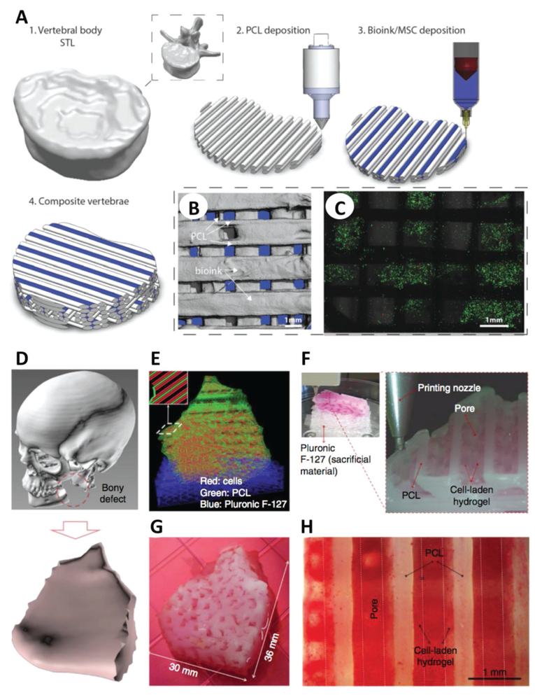
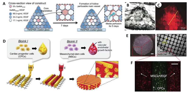
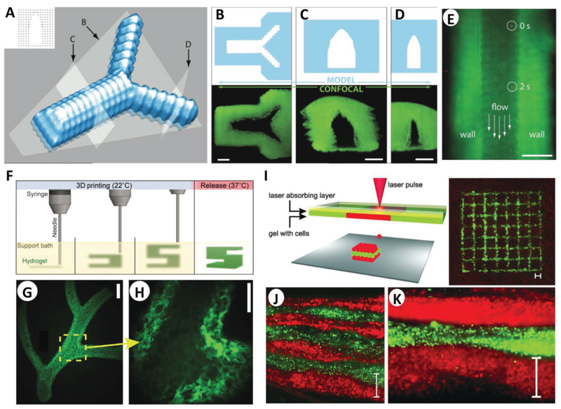
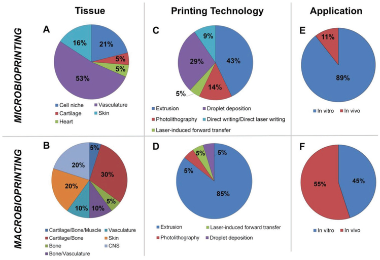
The recapitulation of human anatomy and physiology is critical for organ regeneration. Due to this fundamental requirement, bioprinting holds great promise in tissue engineering and regenerative medicine due to the possibility of fabricating complex scaffolds that host cells and biochemical cues in a physiologically relevant fashion. The ever-growing research in this field has been proceeding along two different, yet complementary, routes: on the one hand, the development of bioprinting to fabricate large tissue surrogates for transplantation purposes in vivo (macrobioprinting), and on the other the spread of bioprinting-based miniaturized systems to model the tissue microenvironment in vitro (microbioprinting). The latest advances in both macro- and microbioprinting are reviewed, emphasizing their impact on specific areas of tissue engineering. Additionally, a critical comparison of macro- versus microbioprinting is presented together with advantages and limitations of each approach. Ultimately, findings obtained both at the macro-and microscale are expected to provide a deeper insight in tissue biology and offer clinically relevant solutions for organ regeneration.

Keywords: bioprinting; complex tissues; organ-on-a-chip; regenerative medicine; tissue engineering.


Conflict of interest statement

Conflict of Interest The authors declare no conflict of interest.

Figures

Figure 1.
Figure 1.
Macrobioprinting of human vertebrae and mandible. A) The geometry of a human vertebra was scanned and bioprinted via extrusion of PCL and MSC-laden alginate/GelMA in an orthogonal fashion. B,C) Microcomputed tomography (B) and live/dead imaging (C) of cells demonstrated the distribution of materials/cells (live cells in green in (C)) within the macrobioprinted vertebrae. A–C) Adapted with permission.[24] Copyright 2016, Wiley-VCH. D–G) Patient data of a mandible defect (D) were used to develop a CAD model for a scaffold (E), which was then bioprinted by depositing alternate fibers of PCL (green), Pluronic F-127 (blue) and cell-laden hydrogel (red) (F,G). H) Alizarin Red staining confirmed the osteogenic differentiation of stem cells in the printed construct. D–H) Adapted by permission.[31] Copyright 2016, Macmillan Publishers Ltd. Scale bars in (B), (C), and (H) are 1 mm.
Figure 2.
Figure 2.
Macrobioprinting of vascularized bone niche and cardiac patch. A) Vascularized bone constructs were fabricated as pyramidal constructs hosting a perfusable vascular lumen lined with HUVECs (pink) and array of hMSCs-laden VEGF-functionalized GelMA fibers with different mechanical strengths. B) Imaging of cross section of the resulting scaffold. C) Imaging of cross-sectional fluorescence gradient to show different chemical functionalization of bioprinted fibers. Scale bars in (B) and (C) are 500 μm. A–C) Adapted with permission.[36] Copyright 2017, Wiley-VCH. D) Bioprinting design of prevascularized cardiac patches, E) where a PCL layer provides structural support for F) two cell-laden bioinks. Scale bar in (E) (left) is 1 mm, while the scale bar in (F) is 200 μm. D–F) Adapted with permission.[39] Copyright 2017, Elsevier.
Figure 3.
Figure 3.
Microbioprinting of vasculature and skin models. A) Rendering of the microvasculature model developed by Pataky et al. B–D) Slices of the model and confocal images according to the slicing planes labeled in (A). E) Composite image showing perfusion of the alginate microchannel with fluorescent beads flowing at 0 and 2 s. The scale bars in (B)–(E) represent 200 μm. A–E) Adapted with permission.[58] Copyright 2012, Wiley-VCH. F) Schematic of the FRESH process developed by Feinberg and co-workers, where a hydrogel (green) is extruded and cross-linked in a gelatin slurry (yellow). Upon completion, the system is heated to 37 °C to melt the gelatin and release the scaffold. G) Example of arterial trees printed in fluorescent alginate (green) via the FRESH method, H) where a stable lumen was formed with and defined vessel wall <1 mm thick. Scale bars are 2.5 and 1 mm in (G) and (H), respectively. F–H) Adapted with permission.[62] Copyright 2015, American Association for the Advancement of Science. I) Schematic of the microbioprinting setup used by Koch et al. (left), where the pressure of a laser-induced vapor bubble propels a cell-laden hydrogel and yields a micropatterned grid structure (right) of fibroblasts (green) and keratinocytes (red). J) Histology image showing seven alternating layers of red and green keratinocytes, where each colored layer consists of four microbioprinted sublayers. Scale bar is 500 μm in (I)–(K). I–K) Adapted with permission.[22] Copyright 2012, Wiley-VCH.
Figure 4.
Figure 4.
Macro- versus microbioprinting. Graphical comparison of the findings in macro- and microbioprinting presented in Sections 2 and 3. Quantitative comparison focused on: A,B) the type of tissues that was bioprinted, C,D) the bioprinting technologies used, and E,F) the application stage in which the constructs were used. A,B) Microbioprinting approaches make use of soft hydrogel as bioink, limiting therefore their applicability to soft-tissue modeling, particularly vasculature and skin. Conversely, macrobioprinting strategies have been mainly leveraged for musculoskeletal TE applications, due to the possibility of combining materials with a wide range of mechanical properties. C,D) A wide range of technologies has been used for tissue modeling via microbioprinting, while macrobioprinting vastly relies on extrusion-based bioprinting to fabricate large constructs in a short amount of time. E,F) As per our definition, microbioprinting strategies focus on the development of highly detailed tissue models, understanding the interaction between cells and their tissue-specific niches, and are thus mainly applied for in vitro studies (89%). A small portion of these studies have been extended into in vivo stages (11%),[57,67] to further assess microprinted features in living environments. The aim of macrobioprinting approaches is to develop viable tissue constructs and the majority has already reached in vivo testing stage (55%) after initial in vitro testing. Despite 45% of applications having not been tested in vivo, they are purposely developed for tissue reconstruction.
Figure 5.
Figure 5.
Acellular micro-3D printing. A,B) Scanning electron microscopy images of pentaerythritol tetraacrylate (PETTA) processed by direct laser writing (A), which was developed by Greiner et al. to investigate cell invasion and morphology (nuclei in red, F-actin in green, and scaffold in white) (B). A,B) Adapted with permission.[72] Copyright 2014, Elsevier. C) Image of microwave sintered tricalcium phosphate (TCP) and Sr-Mg-doped TCP scaffolds, and D) an SEM image of the TCP scaffold. C,D) Adapted with permission.[71] Copyright 2013, The Royal Society of Chemistry.

Similar articles

Cited by

References

    1. Zhang YS, Yue K, Aleman J, Mollazadeh-Moghaddam K, Bakht SM, Yang J, Jia W, Dell’Erba V, Assawes P, Shin SR, Dokmeci MR, Oklu R, Khademhosseini A, Ann. Biomed. Eng 2017, 45, 148. - PMC - PubMed
    1. Ozbolat IT, Peng W, Ozbolat V, Drug Discovery Today 2016, 21, 1257. - PubMed
    1. V Murphy S, Atala A, Nat. Biotechnol 2014, 32, 773. - PubMed
    1. Derby B, Science 2012, 338, 921. - PubMed
    1. Knowlton S, Yenilmez B, Tasoglu S, Trends Biotechnol. 2016, 34, 685. - PubMed

LinkOut - more resources