

Keratoconus severity identification using unsupervised machine learning
- PMID: 30399144
- PMCID: PMC6219768
- DOI: 10.1371/journal.pone.0205998
Keratoconus severity identification using unsupervised machine learning
Abstract
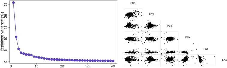
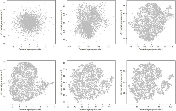
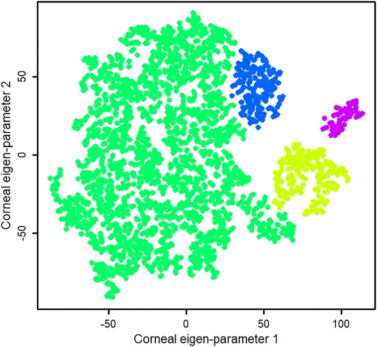
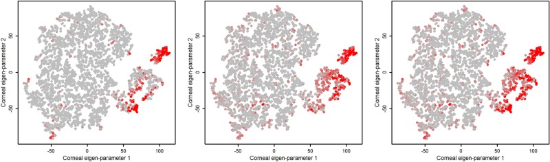
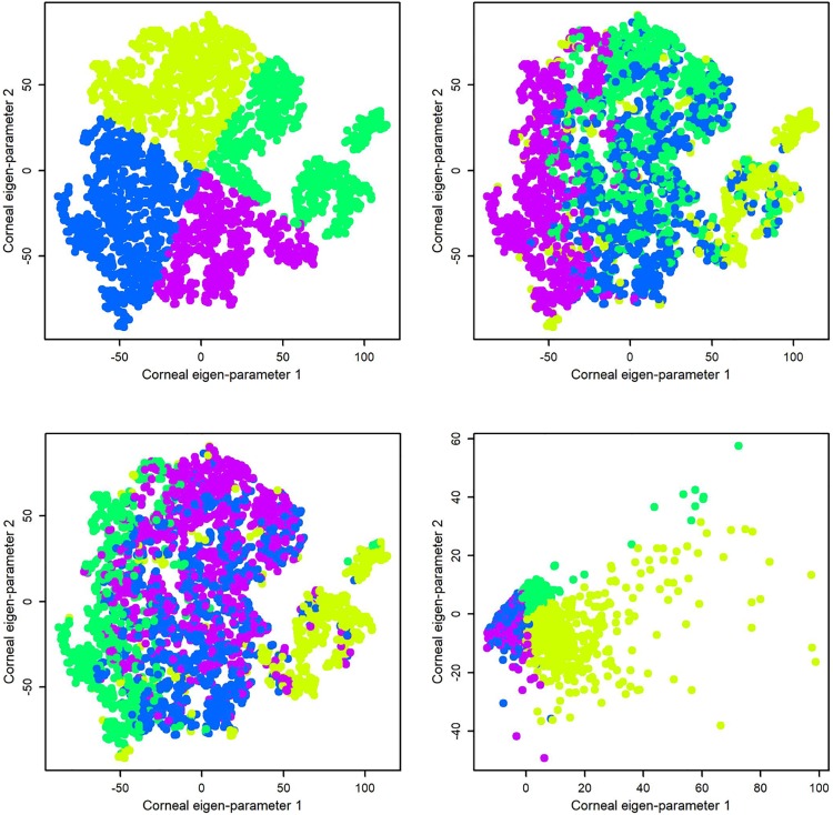
We developed an unsupervised machine learning algorithm and applied it to big corneal parameters to identify and monitor keratoconus stages. A big dataset of corneal swept source optical coherence tomography (OCT) images of 12,242 eyes acquired from SS-1000 CASIA OCT Imaging Systems in multiple centers across Japan was assembled. A total of 3,156 eyes with valid Ectasia Status Index (ESI) between zero and 100% were selected for the downstream analysis. Four hundred and twenty corneal topography, elevation, and pachymetry parameters (excluding ESI Keratoconus indices) were selected. The algorithm included three major steps. 1) Principal component analysis (PCA) was used to linearly reduce the dimensionality of the input data from 420 to eight significant principal components. 2) Manifold learning was used to further reducing the selected principal components nonlinearly to two eigen-parameters. 3) Finally, a density-based clustering was applied to the eigen-parameters to identify eyes with keratoconus. Visualization of clusters in 2-D space was used to validate the quality of learning subjectively and ESI was used to assess the accuracy of the identified clusters objectively. The proposed method identified four clusters; I: a cluster composed of mostly normal eyes (224 eyes with ESI equal to zero, 23 eyes with ESI between five and 29, and nine eyes with ESI greater than 29), II: a cluster composed of mostly healthy eyes and eyes with forme fruste keratoconus (1772 eyes with ESI equal to zero, 698 eyes with ESI between five and 29, and 117 eyes with ESI greater than 29), III: a cluster composed of mostly eyes with mild keratoconus stage (184 eyes with ESI greater than 29, 74 eyes with ESI between five and 29, and 6 eyes with ESI equal to zero), and IV: a cluster composed of eyes with mostly advanced keratoconus stage (80 eyes had ESI greater than 29 and 1 eye had ESI between five and 29). We found that keratoconus status and severity can be well identified using unsupervised machine learning algorithms along with linear and non-linear corneal data transformation. The proposed method can better identify and visualize the keratoconus stages.
Conflict of interest statement
The authors have declared that no competing interests exist.
Figures
References
-
- Rabinowitz YS. Keratoconus. Surv Ophthalmol. 1998;42(4):297–319. . - PubMed
-
- Maeda N, Klyce SD, Smolek MK, Thompson HW. Automated keratoconus screening with corneal topography analysis. Invest Ophthalmol Vis Sci. 1994;35(6):2749–57. . - PubMed
-
- Smolek MK, Klyce SD. Current keratoconus detection methods compared with a neural network approach. Invest Ophthalmol Vis Sci. 1997;38(11):2290–9. . - PubMed
Publication types
MeSH terms
LinkOut - more resources
Full Text Sources
Other Literature Sources
