Review
. 2018 Nov 5;28(21):R1266-R1281.
doi: 10.1016/j.cub.2018.08.034.

Towards a Unified Model of SMC Complex Function

Affiliations
Review

Towards a Unified Model of SMC Complex Function

Markus Hassler et al. Curr Biol. .

Abstract

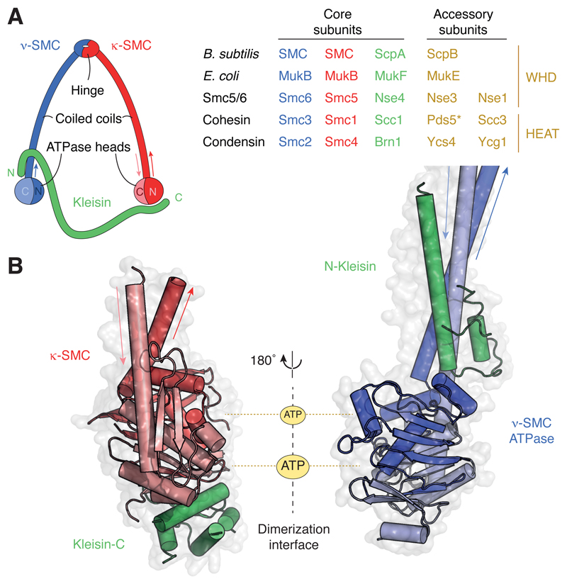
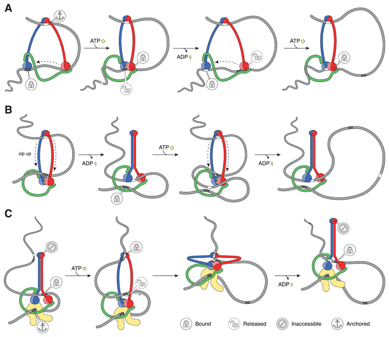
Protein complexes built of structural maintenance of chromosomes (SMC) and kleisin subunits, including cohesin, condensin and the Smc5/6 complex, are master organizers of genome architecture in all kingdoms of life. How these large ring-shaped molecular machines use the energy of ATP hydrolysis to change the topology of chromatin fibers has remained a central unresolved question of chromosome biology. A currently emerging concept suggests that the common principle that underlies the essential functions of SMC protein complexes in the control of gene expression, chromosome segregation or DNA damage repair is their ability to expand DNA into large loop structures. Here, we review the current knowledge about the biochemical and structural properties of SMC protein complexes that might enable them to extrude DNA loops and compare their action to other motor proteins and nucleic acid translocases. We evaluate the currently predominant models of active loop extrusion and propose a detailed version of a 'scrunching' model, which reconciles much of the available mechanistic data and provides an elegant explanation for how SMC protein complexes fulfill an array of seemingly diverse tasks during the organization of genomes.


Figures

Figure 1
Figure 1. Architecture and composition of SMC complexes.
(A) Ring-like architecture created by the association of the SMC and kleisin subunits and subunit composition of common prokaryotic (top two) and eukaryotic (bottom three; S. cerevisiae protein names) SMC complexes. *Note that Pds5 associates with the other cohesin subunits transiently and might hence not be a stoichiometric subunit of cohesin complexes. (B) Structure models of a κ-SMC ATPase head domain bound to the WHD located at the C terminus of the kleisin subunit (pdb: 1w1w; S. cerevisiae cohesin Smc1–Scc1) and of a ν-SMC ATPase head domain bound to the helical domain located at the N terminus of the kleisin subunit (pdb: 4ux3; S. cerevisiae cohesin Smc3–Scc1).
Figure 2
Figure 2. Principles of DNA loop extrusion.
(A) SMC rings might capture the bases of DNA loops either in a topological manner (which requires ring opening for DNA entry or exit) or a pseudo-topological manner (which does not require ring opening for DNA entry). (B) Loop Extrusion by external motors. The transcription machinery might push loops to the 3’ end of actively transcribed genes, thereby enlarging loops. (C) Motor-free models of loop extrusion. The continuous loading of SMC complexes at a specific site creates osmotic pressure that pushes already loaded rings along the DNA until they encounter a barrier (boundary element), where they accumulate and dissociate. (D) SMC complexes as DNA motors. Loop Extrusion could be driven by SMC dimers (handcuffs) that reel in DNA symmetrically from both sides or by individual SMC complexes that anchor DNA at one side and reel in DNA from the other. Symmetric loop extrusion could alternatively be achieved if individual complexes repeatedly switch strands or if two complexes that each anchor DNA assemble in a head-to-head orientation, producing two loops as they reel in DNA from opposite sides. (E) Symmetric loop extrusion until halted by the encounter of boundary elements reproduces TAD formation. Asymmetric loop extrusion is expected to produce ‘stripes’ in Hi-C contact maps and might lead to gaps that are not folded into loops.
Figure 3
Figure 3. Possible DNA binding sites in SMC ATPase head and hinge domains.
(A) Structure model of DNA bound at the shallow surface of the dimerized Methanocaldococcus jannaschii Rad50 ATPase head domains (pdb: 5f3w) and of DNA bound to the coiled coil of one of the Thermotoga maritima Rad50 ATPase head domains (pdb: 4w9m) projected onto the M. jannaschii structure. (B) Structure model of the dimerized T. maritima SMC hinge domain (pdb: 1gxl). Colors represent positive (blue; +5 keT) or negative (red; –5 keT) electrostatic surface potential values.
Figure 4
Figure 4. Conformational changes in SMC proteins.
(A) Models of ATP-bound open (ring-shaped) and ATP-free closed (rod-shaped) SMC dimers and conformational transitions of condensin SMC subunits observed in AFM images. (B) Structure models of the ATP-bound NBD of the Staphylococcus aureus Sav1866 ABC transporter ATPase (pdb: 2onj) and of the Geobacillus stearothermophilus SMC ATPase head domain (pdb: 5h68). (C) Close-up view of the Q-loop conformation in structures of the ATP-free monomeric B. subtilis SMC ATPase head domain (light grey; pdb: 5h67) and the ATP-dimerized B. subtilis SMC ATPase head domain (dark grey; pdb: 5xg3) and side-views of the coiled-coil orientations in both structures. (D) Structural models of the Pyrococcus furiosus SMC hinge domain in the closed coiled-coil conformation (pdb: 4rsj), of the T. maritima SMC hinge domain in the open coiled-coil conformation with both hinge interfaces associated (pdb: 1gxl) and of the G. stearothermophilus SMC hinge domain in the open coiled-coil conformation with one hinge interface dissociated (pdb: 5h69).
Figure 5
Figure 5. Different models of active SMC-driven Loop Extrusion mechanism.
(A) Sequential walking model. (B) DNA pumping model. (C) Extended scrunching model. See main text for details. Note that the sequential walking model makes no assumptions about the SMC coiled coil conformations, whereas the pumping model assumes stiff coiled coils that are under tension when bent open. The extended scrunching models postulates that SMC coiled coils alternate between stiff and relaxed states.

References

    1. Cobbe N, Heck MMS. The evolution of ATPase activity in SMC proteins. Proteins. 2006;63:685–96. - PubMed
    1. Gruber S. MukBEF on the march: taking over chromosome organization in bacteria? Mol Microbiol. 2011;81:855–859. - PubMed
    1. Uhlmann F. SMC complexes: from DNA to chromosomes. Nat Rev Mol Cell Biol. 2016;17:399–412. - PubMed
    1. van Ruiten MS, Rowland BD. SMC Complexes: Universal DNA Looping Machines with Distinct Regulators. Trends Genet. 2018;34:477–487. - PubMed
    1. Bürmann F, Basfeld A, Vazquez Nunez R, Diebold-Durand M-L, Wilhelm L, Gruber S. Tuned SMC Arms Drive Chromosomal Loading of Prokaryotic Condensin. Mol Cell. 2017;65:861–869. - PMC - PubMed

Publication types

MeSH terms

Substances

LinkOut - more resources