

Crocin Ameliorates Atopic Dermatitis Symptoms by down Regulation of Th2 Response via Blocking of NF-κB/STAT6 Signaling Pathways in Mice
- PMID: 30400140
- PMCID: PMC6266819
- DOI: 10.3390/nu10111625
Crocin Ameliorates Atopic Dermatitis Symptoms by down Regulation of Th2 Response via Blocking of NF-κB/STAT6 Signaling Pathways in Mice
Abstract
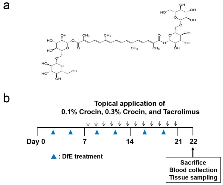
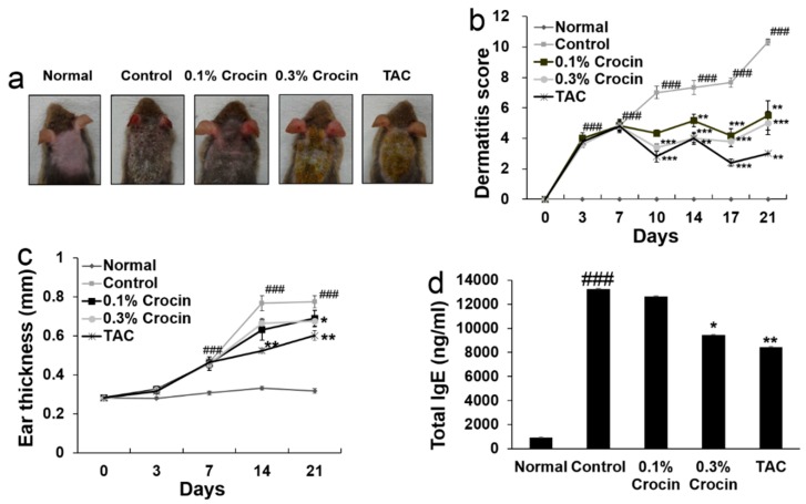
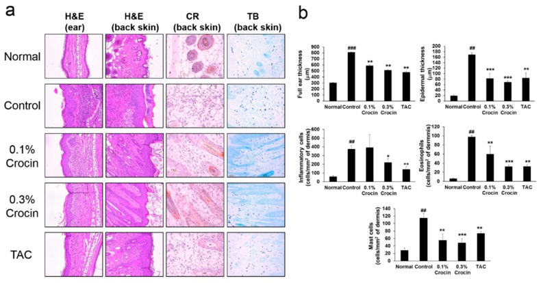
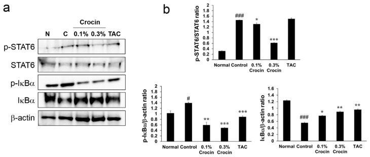
Crocin, a major constituent of Gardenia jasminoides, is a natural colorant carotenoid compound that has been reported to have anti-inflammatory effects. This study investigated the therapeutic effects of crocin on mice with atopic dermatitis induced by Dermatophagoides farinae crude extract, which is a common environmental allergen in house dust that causes atopic dermatitis in humans. Crocin application ameliorated Dermatophagoides farinae crude extract-induced atopic dermatitis symptoms by inhibiting the dermatitis severity score, ear thickness, and serum immunoglobulin E levels in NC/Nga mice. The increases in epidermal thickness and dermal inflammatory cells (eosinophil and mast cells) infiltrations observed on the dorsal back skin of atopic dermatitis control mice were inhibited in a dose-dependent manner by topical application of crocin in atopic dermatitis treatment mice. Crocin inhibited the Dermatophagoides farinae crude extract-induced increase of thymus and activation-regulated chemokines, interleukin (IL)-4, and IL-13 on the dorsal skin of mice. Crocin also inhibited Dermatophagoides farinae crude extract-induced activation of nuclear factor-κB (NF-κB) and signal transducer and activator of transcription (STAT) 6. These results show that crocin ameliorates atopic dermatitis symptoms by down regulation of the Th2 cells-mediated immune response via blocking of NF-κB/STAT6 signaling pathways.
Keywords: Gardenia jasminoides; Immunoglobulin E; NC/Nga; eosinophils; thymus and activation-regulated chemokine.
Conflict of interest statement
The authors declare no conflict of interest.
Figures
Similar articles
-
Gardenia jasminoides extract without crocin improved atopic dermatitis-like skin lesions via suppression of Th2-related cytokines in Dfe-induced NC/Nga mice.J Ethnopharmacol. 2019 Sep 15;241:112015. doi: 10.1016/j.jep.2019.112015. Epub 2019 Jun 4. J Ethnopharmacol. 2019. PMID: 31173875
-
The Gardenia jasminoides extract and its constituent, geniposide, elicit anti-allergic effects on atopic dermatitis by inhibiting histamine in vitro and in vivo.J Ethnopharmacol. 2014 Oct 28;156:33-40. doi: 10.1016/j.jep.2014.07.060. Epub 2014 Aug 19. J Ethnopharmacol. 2014. PMID: 25153023
-
Topical application of an ethanol extract prepared from Illicium verum suppresses atopic dermatitis in NC/Nga mice.J Ethnopharmacol. 2012 Oct 31;144(1):151-9. doi: 10.1016/j.jep.2012.08.042. Epub 2012 Sep 3. J Ethnopharmacol. 2012. PMID: 22971899
-
[Therapeutic agents of today and the future for atopic dermatitis].Nihon Yakurigaku Zasshi. 2006 Dec;128(6):411-5. doi: 10.1254/fpj.128.411. Nihon Yakurigaku Zasshi. 2006. PMID: 17167215 Review. Japanese. No abstract available.
-
Crocin: A fighter against inflammation and pain.Food Chem Toxicol. 2020 Sep;143:111521. doi: 10.1016/j.fct.2020.111521. Epub 2020 Jul 5. Food Chem Toxicol. 2020. PMID: 32640351 Review.
Cited by
-
SNAP25 protects primary cortical neurons from hypoxic-ischemic injury associated with CREB signal.Ibrain. 2021 Mar 28;7(1):1-11. doi: 10.1002/j.2769-2795.2021.tb00058.x. eCollection 2021 Mar. Ibrain. 2021. PMID: 37786874 Free PMC article.
-
Perspectives of herbs and their natural compounds, and herb formulas on treating diverse diseases through regulating complicated JAK/STAT signaling.Front Pharmacol. 2022 Oct 17;13:993862. doi: 10.3389/fphar.2022.993862. eCollection 2022. Front Pharmacol. 2022. PMID: 36324680 Free PMC article. Review.
-
Gardenia Jasminoides Ameliorates Antibiotic-Associated Aggravation of DNCB-Induced Atopic Dermatitis by Restoring the Intestinal Microbiome Profile.Nutrients. 2021 Apr 18;13(4):1349. doi: 10.3390/nu13041349. Nutrients. 2021. PMID: 33919521 Free PMC article.
-
Suppression of SAMSN1 contributes to neuroprotection in neonatal rats suffering from hypoxic-ischemic encephalopathy injury.Ibrain. 2022 Nov 12;9(1):3-12. doi: 10.1002/ibra.12078. eCollection 2023 Spring. Ibrain. 2022. PMID: 37786523 Free PMC article.
-
Gardenia jasminoides Attenuates Allergic Rhinitis-Induced Inflammation by Inhibiting Periostin Production.Pharmaceuticals (Basel). 2021 Sep 28;14(10):986. doi: 10.3390/ph14100986. Pharmaceuticals (Basel). 2021. PMID: 34681210 Free PMC article.
References
MeSH terms
Substances
Grants and funding
LinkOut - more resources
Full Text Sources
Research Materials
Miscellaneous
