Review
. 2018 Nov 2;19(11):3447.
doi: 10.3390/ijms19113447.

PGC-1α as a Pivotal Factor in Lipid and Metabolic Regulation

Affiliations
Review

PGC-1α as a Pivotal Factor in Lipid and Metabolic Regulation

Ching-Feng Cheng et al. Int J Mol Sci. .

Abstract

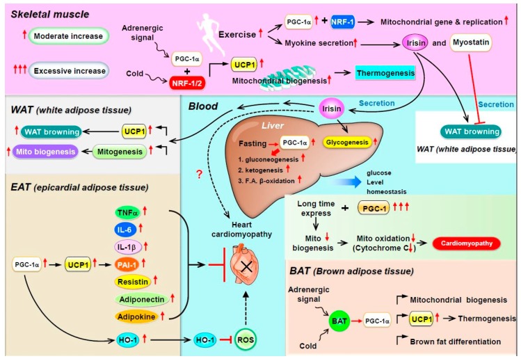
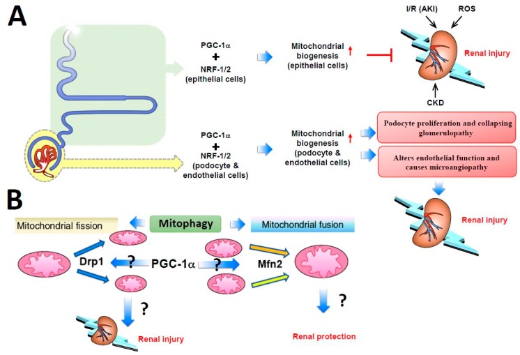
Traditionally, peroxisome proliferator-activated receptor γ coactivator 1α (PGC-1α), a 91 kDa transcription factor, regulates lipid metabolism and long-chain fatty acid oxidation by upregulating the expression of several genes of the tricarboxylic acid cycle and the mitochondrial fatty acid oxidation pathway. In addition, PGC-1α regulates the expression of mitochondrial genes to control mitochondria DNA replication and cellular oxidative metabolism. Recently, new insights showed that several myokines such as irisin and myostatin are epigenetically regulated by PGC-1α in skeletal muscles, thereby modulating systemic energy balance, with marked expansion of mitochondrial volume density and oxidative capacity in healthy or diseased myocardia. In addition, in our studies evaluating whether PGC-1α overexpression in epicardial adipose tissue can act as a paracrine organ to improve or repair cardiac function, we found that overexpression of hepatic PGC-1α increased hepatic fatty acid oxidation and decreased triacylglycerol storage and secretion in vivo and in vitro. In this review, we discuss recent studies showing that PGC-1α may regulate mitochondrial fusion⁻fission homeostasis and affect the renal function in acute or chronic kidney injury. Furthermore, PGC-1α is an emerging protein with a biphasic role in cancer, acting both as a tumor suppressor and a tumor promoter and thus representing a new and unresolved topic for cancer biology studies. In summary, this review paper demonstrates that PGC-1α plays a central role in coordinating the gene expression of key components of mitochondrial biogenesis and as a critical metabolic regulator in many vital organs, including white and brown adipose tissue, skeletal muscle, heart, liver, and kidney.

Keywords: PGC-1α; adipose tissue; metabolic homeostasis; mitochondria.


Conflict of interest statement

The authors declare no conflict of interest.

Figures

Figure 1
Figure 1
Schematic description of peroxisome proliferator-activated receptor gamma coactivator-1α (PGC-1α) function in various organs. Traditionally, in brown adipocytes (BAT) and white adipocytes (WAT), mitochondrial biogenesis and BAT gene expression are regulated by PGC-1α. Adrenergic stimulation and lower temperature trigger signaling cascades, including the upregulation of UCP-1 level, thereby resulting in body thermogenesis. In skeletal muscle and WAT, the transcriptional activity of PGC-1α is responsible for the expression of gene networks that control glucose uptake, glycolysis, fatty acid (FA) oxidation, tricarboxylic acid cycle, oxidative phosphorylation (OXPHOS), mitochondrial biogenesis, and protein uncoupling. Therefore, increasing exercise will increase mitochondrial gene biogenesis and secretion of myokines (such as irisin), which results in WAT browning and liver gluconeogenesis to prevent obesity and insulin resistance. In epicardial adipose tissue (EAT), increased heme oxygenase 1 (HO-1) expression depends on the PGC-1α–UCP-1 axis activity, which then decreases free radicals and reactive oxygen species (ROS) production, thus reducing cardiomyopathy. However, whether increased expression of cytokines such as TNF-α, IL-6, or adipokines by the PGC-1α–UCP-1 axis can reduce cardiomyopathy or not is still unclear.
Figure 2
Figure 2
Schematic description of PGC-1α function in renal homeostasis. (A) PGC-1α associated with nuclear respiratory factors 1 and 2 (NRF-1/2) has a protective role in renal epithelial cells, including the proximal convoluted tubule, loop of Henle, and distal convoluted tubule, during renal injury by increasing mitochondrial biogenesis in epithelial cells. Bowman’s capsule, podocytes, and endothelial cells have a narrow PGC-1α tolerance. Increased PGC-1α levels in podocytes induce podocyte proliferation and collapsing glomerulopathy development, whereas increased PGC1-α in endothelial cells alters endothelial function and causes microangiopathy, thereby resulting in renal injury. (B) The role of mitochondrial fusion and fission in mitophagy. Mitochondrial fusion is promoted by the Mfn2 gene, whereas Drp1 promotes mitochondrial fission. Increased Drp1 and decreased Mfn2 expression exacerbates tubular damage, thereby contributing to kidney disease; however, studies have shown opposite results and inconsistencies. Whether PGC-1α transcriptionally regulates Drp1 and Mfn2 requires further research.

References

    1. Besseiche A., Riveline J.P., Gautier J.F., Breant B., Blondeau B. Metabolic roles of PGC-1alpha and its implications for type 2 diabetes. Diabetes Metab. 2015;41:347–357. doi: 10.1016/j.diabet.2015.02.002. - DOI - PubMed
    1. Puigserver P., Wu Z., Park C.W., Graves R., Wright M., Spiegelman B.M. A cold-inducible coactivator of nuclear receptors linked to adaptive thermogenesis. Cell. 1998;92:829–839. doi: 10.1016/S0092-8674(00)81410-5. - DOI - PubMed
    1. Popov D.V., Lysenko E.A., Kuzmin I.V., Vinogradova V., Grigoriev A.I. Regulation of PGC-1alpha Isoform Expression in Skeletal Muscles. Acta Nat. 2015;7:48–59. - PMC - PubMed
    1. Lin J., Handschin C., Spiegelman B.M. Metabolic control through the PGC-1 family of transcription coactivators. Cell Metab. 2005;1:361–370. doi: 10.1016/j.cmet.2005.05.004. - DOI - PubMed
    1. Hatazawa Y., Senoo N., Tadaishi M., Ogawa Y., Ezaki O., Kamei Y., Miura S. Metabolomic Analysis of the Skeletal Muscle of Mice Overexpressing PGC-1alpha. PLoS ONE. 2015;10:e0129084. doi: 10.1371/journal.pone.0129084. - DOI - PMC - PubMed

Substances

LinkOut - more resources