. 2018 Nov 2;7(11):410.
doi: 10.3390/jcm7110410.

Mechanical Stretching Simulates Cardiac Physiology and Pathology through Mechanosensor Piezo1

Affiliations

Mechanical Stretching Simulates Cardiac Physiology and Pathology through Mechanosensor Piezo1

Tzyy-Yue Wong et al. J Clin Med. .

Abstract

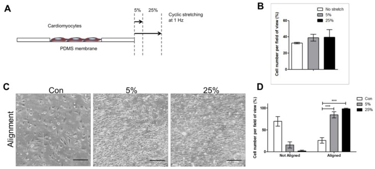
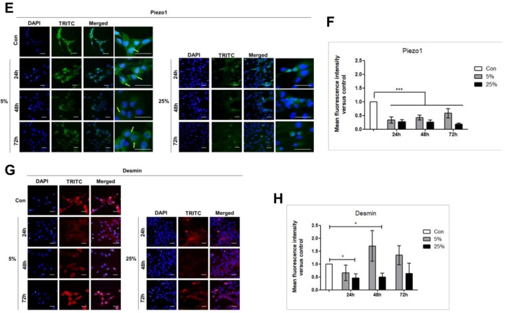
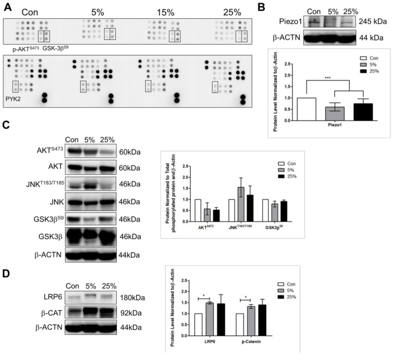
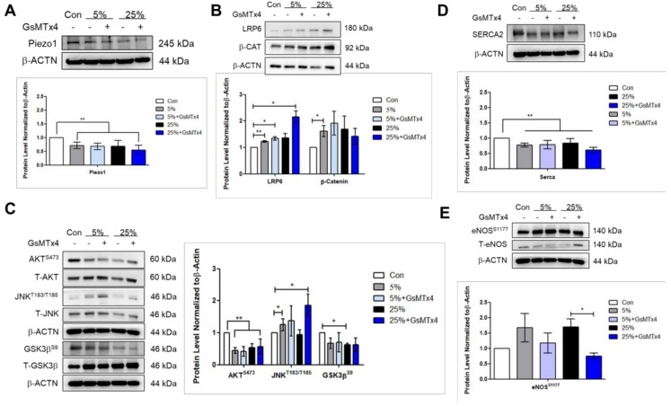
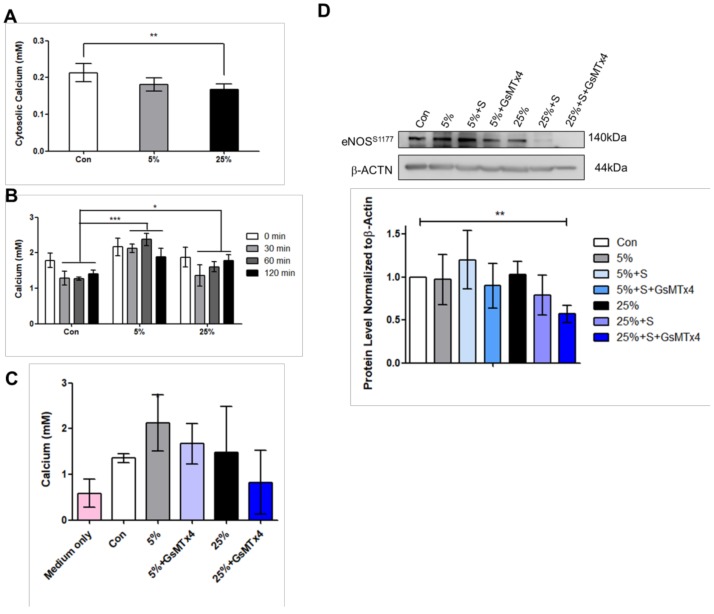
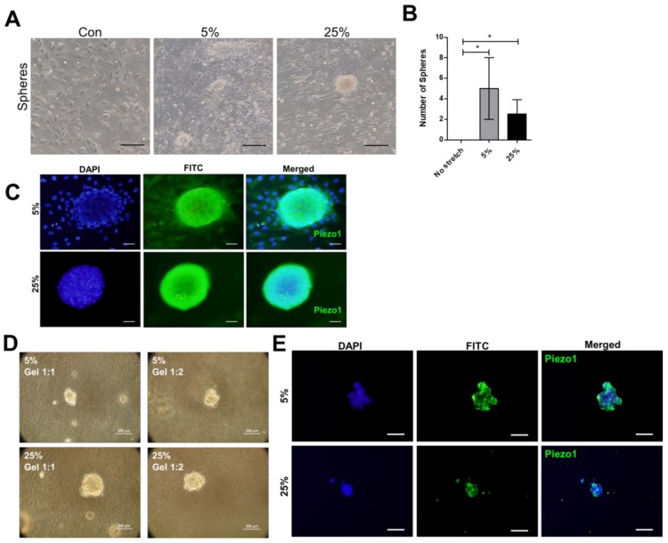
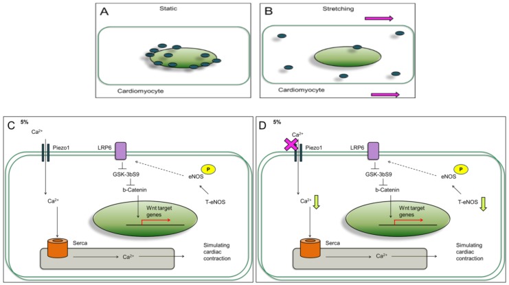
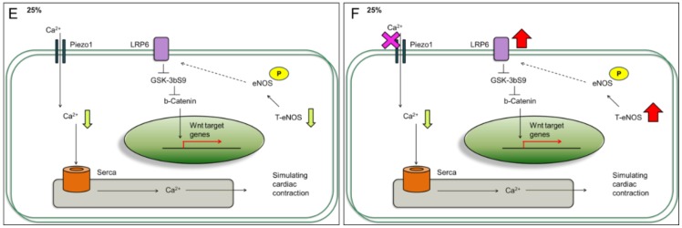
The dynamics of a living body enables organs to experience mechanical stimulation at cellular level. The human cardiomyocytes cell line provides a source for simulating heart dynamics; however, a limited understanding of the mechanical stimulation effect on them has restricted potential applications. Here, we investigated the effect of mechanical stimulation on the cardiac function-associated protein expressions in human cardiomyocytes. Human cardiomyocyte cell line AC16 was subjected to different stresses: 5% mild and 25% aggressive, at 1 Hz for 24 h. The stretched cardiomyocytes showed down-regulated Piezo1, phosphorylated-Ak transforming serine473 (P-AKTS473), and phosphorylated-glycogen synthase kinase-3 beta serine9 P-GSK3βS9 compared to no stretch. In addition, the stretched cardiomyocytes showed increased low-density lipoprotein receptor-related protein 6 (LRP6), and phosphorylated-c-Jun N-terminal kinase threonine183/tyrosine185 (P-JNKT183/Y185). When Piezo inhibitor was added to the cells, the LRP6, and P-JNKT183/Y185 were further increased under 25%, but not 5%, suggesting that higher mechanical stress further activated the wingless integrated-(Wnt)-related signaling pathway when Piezo1 was inhibited. Supporting this idea, when Piezo1 was inhibited, the expression of phosphorylated-endothelial nitric oxide synthase serine1177 (P-eNOSS1177) and release of calcium ions were reduced under 25% compared to 5%. These studies demonstrate that cyclic mechanical stimulation affects cardiac function-associated protein expressions, and Piezo1 plays a role in the protein regulation.

Keywords: Piezo1; cardiomyocytes; mechanical stimulation; stretching.


Conflict of interest statement

The authors declare no conflict of interest.

Figures

Figure 1
Figure 1
Cyclic stretch affects cardiomyocyte alignment and Piezo1 distribution. (A) Illustration showing cardiomyocytes seeded on stretchable polydimethylsiloxane PDMS membrane, stretched in one direction or uniaxial. (B) Cell number was counted after subjected to 5%, and 25% 24 h stretching. (C,D) Cell alignment after 24 h was measured using ImageJ. (E,F) Expressions of the stretch-activated ion channel Piezo1. (G,H) and cardiomyocyte characteristic marker Desmin analyzed by immunofluorescence assay after 24 h, 48 h, and 72 h of stretching. Scale bar = 100 μm. *, p value < 0.05, ***, p value < 0.001.
Figure 1
Figure 1
Cyclic stretch affects cardiomyocyte alignment and Piezo1 distribution. (A) Illustration showing cardiomyocytes seeded on stretchable polydimethylsiloxane PDMS membrane, stretched in one direction or uniaxial. (B) Cell number was counted after subjected to 5%, and 25% 24 h stretching. (C,D) Cell alignment after 24 h was measured using ImageJ. (E,F) Expressions of the stretch-activated ion channel Piezo1. (G,H) and cardiomyocyte characteristic marker Desmin analyzed by immunofluorescence assay after 24 h, 48 h, and 72 h of stretching. Scale bar = 100 μm. *, p value < 0.05, ***, p value < 0.001.
Figure 2
Figure 2
Cyclic stretch stimulates the low-density lipoprotein receptor-related protein 6 LRP6/β-catenin signaling. (A) Cardiomyocytes analyzed by Phospho-kinase array at no stretch (control), 5%, 15%, and 25% 24 h of stretching. (B) Western blot performed to analyze the stretch-activated ion channel Piezo1 protein expression. Validation of the (C) phosphorylated-Ak transforming serine473 (AKTS473), phosphorylated-c-Jun N-terminal kinase threonine183/tyrosine185 (JNKT183/Y185), phosphorylated-glycogen synthase kinase-3 beta serine9 (GSK3βS9), total AKT, JNK, and GSK3β, (D) wingless integrated (Wnt) signaling molecules LRP6, and β-catenin. Data represent at least three independent experiments in SEM ± SD. *, p value < 0.05, ***, p value < 0.001.
Figure 3
Figure 3
Aggressive mechanical stimulation further activates the LRP6/β-catenin signaling when Piezo1 was inhibited. Western blot showing the protein expressions for (A) Piezo1, (B) LRP6, and β-catenin, (C) phosphorylated AKTS473, JNKT183/Y185, GSK3βS9, and total AKT, JNK, and GSK3β. (D,E) Protein level for the calcium ion regulator sarco/endoplasmic reticulum Ca2+ SERCA2, phosphorylated-endothelial nitric oxide synthase serine1177 P-eNOSS1177, and total eNOS. Data represent at least three independent experiments in SEM ± SD. *, p value < 0.05, **, p value < 0.01.
Figure 4
Figure 4
Aggressive mechanical stimulation reduces calcium ion and P-eNOS levels. (A) Cardiomyocytes cell pellets were collected and lysed in lysis buffer after 24 h stretching, cytosolic calcium ion determined by colorimetric detection kit. (B) Calcium ions released into the medium was measured immediately after stretching at 0 min, 30, 60, and 120 min. The medium which was the supernatant were collected simultaneously, absorbance measured at 575 nm. (C) Calcium ion released into the medium was measured after 24 h stretching with Piezo1 inhibited (Piezo1 inhibitor GsMTx4) at 5%, and 25%. (D) After 21 h of stretching, the cardiac drug simvastatin, or GsMTx4 was added to the cells, and continued cultured for 3 h before collecting cell lysates at 24 h. Data represent at least three independent experiments in SEM ± SD. *, p value < 0.05, **, p value < 0.01, ***, p value < 0.001.
Figure 5
Figure 5
Expression of the Wnt1 protein in the isopretenol-induced myocardial infarction rat model. (A) Analysis of Wnt1 protein expression in normal rat by immunohistochemistry. (B) Wnt1 protein expression in the isopretenol (ISO)-induced myocardial infarction (MI) rat model (n = 3). (C) Scoring of the tissue slides analyzed by immunohistochemistry: 0 (no staining); 1 (<10% staining); 2 (10–50% staining); 3 (>50% staining). *, p value < 0.05.
Figure 6
Figure 6
Cyclic stretch enhances growth of clustered cells expressing Piezo1 that survive in 3D hydrogel. (A,B) The 24 h stretching formed clustered cells with spherical appearance. Scale bar = 100 μm. (C) Immunofluorescence assay detected Piezo1 in the clustered cardiomyocytes. Scale bar = 20 μm. (D) The clustered cardiomyocytes were transferred to hydrogel, cultured for 7 days with cover medium changed every 2 days. Scale bar = 200 μm. (E) After 7 days, the cells in hydrogel were embedded in paraffin, analyzed for Piezo1 expression by immunohistochemistry. Scale bar = 200 μm. Data represent at least three independent experiments in SEM ± SD. *, p value < 0.05.
Figure 7
Figure 7
The effects of mild and aggressive mechanical stimulations on LRP6, JNK signaling, eNOS production, and calcium ions in cardiomyocytes. (A,B) Piezo1 distribution observed to be different after subjected to mild and aggressive mechanical stimulations. (C) Cardiomyocytes subjected to 5% stretching for 24 h increased the LRP6/β-catenin (Wnt pathway) signaling. The SERCA2 is known to act as calcium transporter on the sarco/endoplasmic reticulum to promote cell contractility. In this study, SERCA was detected in the cardiomyocytes. (D) Piezo1 inhibition reduced calcium release at 5%. (E) At 25%, calcium ion release and T-eNOS protein level were reduced compared to 5%. (F) Piezo1 inhibition at 25% reduced calcium ion release; however, significantly increased LRP6/β-catenin, and JNK.
Figure 7
Figure 7
The effects of mild and aggressive mechanical stimulations on LRP6, JNK signaling, eNOS production, and calcium ions in cardiomyocytes. (A,B) Piezo1 distribution observed to be different after subjected to mild and aggressive mechanical stimulations. (C) Cardiomyocytes subjected to 5% stretching for 24 h increased the LRP6/β-catenin (Wnt pathway) signaling. The SERCA2 is known to act as calcium transporter on the sarco/endoplasmic reticulum to promote cell contractility. In this study, SERCA was detected in the cardiomyocytes. (D) Piezo1 inhibition reduced calcium release at 5%. (E) At 25%, calcium ion release and T-eNOS protein level were reduced compared to 5%. (F) Piezo1 inhibition at 25% reduced calcium ion release; however, significantly increased LRP6/β-catenin, and JNK.

Similar articles

Cited by

References

    1. King T.C. Elsevier’s Integrated Pathology. Mosby; Philadelphia, PA, USA: 2007. Cardiovascular Pathology; pp. 169–195.
    1. Peyronnet R., Nerbonne J.M., Kohl P. Cardiac mechano-gated ion channels and arrhythmias. Circ. Res. 2016;118:311–329. doi: 10.1161/CIRCRESAHA.115.305043. - DOI - PMC - PubMed
    1. Dostal D.E., Feng H., Nizamutdinov D., Golden H.B., Afroze S.H., Dostal J.D., Jacob J.C., Foster D.M., Tong C., Glaser S., et al. Mechanosensing and Regulation of Cardiac Function. J. Clin. Exp. Cardiol. 2014;5:314. - PMC - PubMed
    1. Fiorillo C., Nediani C., Ponziani V., Giannini L., Celli A., Nassi N., Formigli L., Perna A.M., Nassi P. Cardiac volume overload rapidly induces oxidative stress-mediated myocyte apoptosis and hypertrophy. Biochim. Biophys. Acta (BBA)—Mol. Basis Dis. 2005;1741:173–182. doi: 10.1016/j.bbadis.2005.03.015. - DOI - PubMed
    1. Tirziu D., Simons M. Endothelium-driven myocardial growth or nitric oxide at the crossroads. Trends Cardiovasc. Med. 2008;18:299–305. doi: 10.1016/j.tcm.2009.01.002. - DOI - PMC - PubMed