Review
. 2018 Nov 3;19(11):3454.
doi: 10.3390/ijms19113454.

Circular RNAs in Muscle Function and Disease

Affiliations
Review

Circular RNAs in Muscle Function and Disease

Simona Greco et al. Int J Mol Sci. .

Abstract

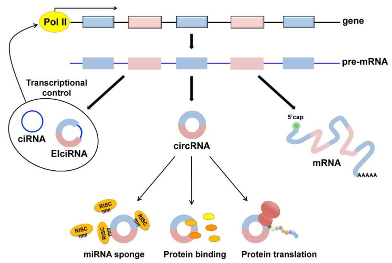
Circular RNAs (circRNAs) are a class of RNA produced during pre-mRNA splicing that are emerging as new members of the gene regulatory network. In addition to being spliced in a linear fashion, exons of pre-mRNAs can be circularized by use of the 3' acceptor splice site of upstream exons, leading to the formation of circular RNA species. In this way, genetic information can be re-organized, increasing gene expression potential. Expression of circRNAs is developmentally regulated, tissue and cell-type specific, and shared across eukaryotes. The importance of circRNAs in gene regulation is now beginning to be recognized and some putative functions have been assigned to them, such as the sequestration of microRNAs or proteins, the modulation of transcription, the interference with splicing, and translation of small proteins. In accordance with an important role in normal cell biology, circRNA deregulation has been reported to be associated with diseases. Recent evidence demonstrated that circRNAs are highly expressed in striated muscle tissue, both skeletal and cardiac, that is also one of the body tissue showing the highest levels of alternative splicing. Moreover, initial studies revealed altered circRNA expression in diseases involving striated muscle, suggesting important functions of these molecules in the pathogenetic mechanisms of both heart and skeletal muscle diseases. The recent findings in this field will be described and discussed.

Keywords: cardiac muscle; circular RNAs; muscle disease; skeletal muscle.


Conflict of interest statement

The authors declare no conflict of interest.

Figures

Figure 1
Figure 1
Putative functions of circRNAs. In addition to the production of messenger RNA (mRNA), back splicing events from the pre-mRNA molecules may lead to the formation of circRNAs. Three types of circRNAs have been identified, exonic (circRNAs), intronic (ciRNAs), and exon-intronic (EIciRNAs). ciRNAs and EIciRNAs have been shown to play a role in the modulation of the transcription of their parental genes (bold arrows). The most abundant exonic circRNAs have been implicated in sponging miRNAs associated with the RNA-induced silencing complex (RISC) through multiple miRNA binding sites in the sequestration of bound proteins and in protein translation through a cap-independent mechanism (narrow arrows).
Figure 2
Figure 2
Strategies to modulate circRNA expression. (1) Plasmids designed to overexpress circRNAs contain the circRNA sequence (colored boxes) flanked by splicing signals (not shown) and intronic sequences including inverted repeats (IR, red dots). IR pairing within the transcribed RNA induces the formation of a secondary structure that facilitates back splicing and circularization. TSS: Transcriptional Start Site; Term: Transcriptional Terminator. Produced circRNAs can then (a) sequester miRNA-RISC complexes to inhibit miRNA action on mRNA targets or (b) interact with RNA-binding proteins (RBPs), sequestering them and preventing their activity; (2) silencing of circRNAs can be achieved through treatment with a siRNA complementary to the back-splice junction sequence (Junction siRNA). This should allow us to target specifically the circRNA, but not the corresponding linear transcript. The siRNA, loaded on the RISC complex, can selectively bind the circRNA and induce its degradation by endonucleolytic cleavage.

Similar articles

Cited by

References

    1. Lamb G.D. Excitation-contraction coupling in skeletal muscle: Comparisons with cardiac muscle. Clin. Exp. Pharmacol. Physiol. 2000;27:216–224. doi: 10.1046/j.1440-1681.2000.03224.x. - DOI - PubMed
    1. Edmondson D.G., Lyons G.E., Martin J.F., Olson E.N. Mef2 gene expression marks the cardiac and skeletal muscle lineages during mouse embryogenesis. Development. 1994;120:1251–1263. - PubMed
    1. Fijnvandraat A.C., Lekanne Deprez R.H., Moorman A.F. Development of heart muscle-cell diversity: A help or a hindrance for phenotyping embryonic stem cell-derived cardiomyocytes. Cardiovasc. Res. 2003;58:303–312. doi: 10.1016/S0008-6363(03)00246-3. - DOI - PubMed
    1. Chal J., Pourquie O. Making muscle: Skeletal myogenesis in vivo and in vitro. Development. 2017;144:2104–2122. doi: 10.1242/dev.151035. - DOI - PubMed
    1. Falcone G., Perfetti A., Cardinali B., Martelli F. Noncoding RNAs: Emerging players in muscular dystrophies. Biomed. Res. Int. 2014 doi: 10.1155/2014/503634. - DOI - PMC - PubMed

MeSH terms

LinkOut - more resources