

Study of Osteocyte Behavior by High-Resolution Intravital Imaging Following Photo-Induced Ischemia
- PMID: 30400346
- PMCID: PMC6278482
- DOI: 10.3390/molecules23112874
Study of Osteocyte Behavior by High-Resolution Intravital Imaging Following Photo-Induced Ischemia
Abstract
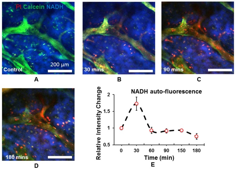
Ischemic injuries and local hypoxia can result in osteocytes dysfunction and play a key role in the pathogenesis of avascular osteonecrosis. Conventional imaging techniques including magnetic resonance imaging (MRI) and computed tomography (CT) can reveal structural and functional changes within bony anatomy; however, characterization of osteocyte behavioral dynamics in the setting of osteonecrosis at the single cell resolution is limited. Here, we demonstrate an optical approach to study real-time osteocyte functions in vivo. Using nicotinamide adenine dinucleotide (NADH) as a biomarker for metabolic dynamics in osteocytes, we showed that NADH level within osteocytes transiently increase significantly after local ischemia through non-invasive photo-induced thrombosis of afferent arterioles followed by a steady decline. Our study presents a non-invasive optical approach to study osteocyte behavior through the modulation of local environmental conditions. Thus it provides a powerful toolkit to study cellular processes involved in bone pathologies in vivo.
Keywords: NADH; osteocyte; osteonecrosis; two-photon.
Conflict of interest statement
The authors declare no conflicts of interest.
Figures
Similar articles
-
Regional differences in oxidative metabolism and mitochondrial activity among cortical bone osteocytes.Bone. 2016 Sep;90:15-22. doi: 10.1016/j.bone.2016.05.011. Epub 2016 May 31. Bone. 2016. PMID: 27260646 Free PMC article.
-
Apoptosis of osteocytes in glucocorticoid-induced osteonecrosis of the hip.J Clin Endocrinol Metab. 2000 Aug;85(8):2907-12. doi: 10.1210/jcem.85.8.6714. J Clin Endocrinol Metab. 2000. PMID: 10946902
-
An electron microscopic study of the changes observed in osteocytes under ischemic conditions.J Orthop Res. 1989;7(1):12-21. doi: 10.1002/jor.1100070103. J Orthop Res. 1989. PMID: 2908902
-
Role of apoptosis in glucocorticoid-induced osteoporosis and osteonecrosis.Crit Rev Eukaryot Gene Expr. 2003;13(2-4):221-35. Crit Rev Eukaryot Gene Expr. 2003. PMID: 14696969 Review.
-
Update on the correlation between mitochondrial function and osteonecrosis of the femoral head osteocytes.Redox Rep. 2025 Dec;30(1):2491846. doi: 10.1080/13510002.2025.2491846. Epub 2025 Apr 18. Redox Rep. 2025. PMID: 40249372 Free PMC article. Review.
Cited by
-
Osteocytes and Cancer.Curr Osteoporos Rep. 2021 Dec;19(6):616-625. doi: 10.1007/s11914-021-00712-9. Epub 2021 Nov 13. Curr Osteoporos Rep. 2021. PMID: 34773212 Free PMC article. Review.
References
-
- Mankin H.J. Nontraumatic necrosis of bone (osteonecrosis) N. Engl. J. Med. 1992;326:1473–1479. - PubMed
MeSH terms
Substances
LinkOut - more resources
Full Text Sources
Medical
