Review
. 2017 Aug 19;8(8):255.
doi: 10.3390/mi8080255.

Biomaterials Meet Microfluidics: From Synthesis Technologies to Biological Applications

Affiliations
Review

Biomaterials Meet Microfluidics: From Synthesis Technologies to Biological Applications

Jingyun Ma et al. Micromachines (Basel). .

Abstract

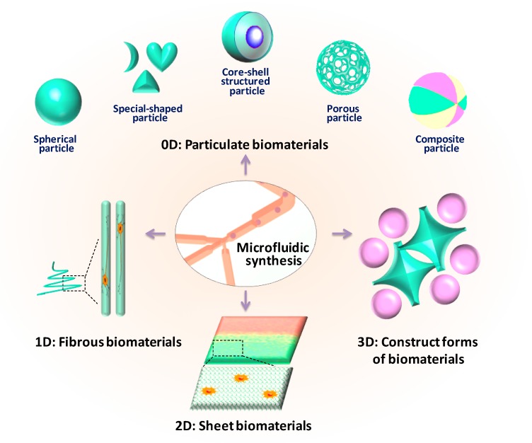
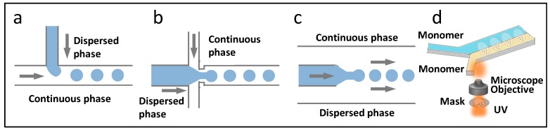
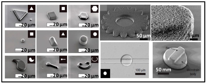
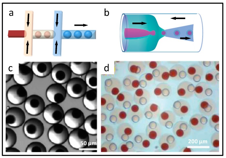
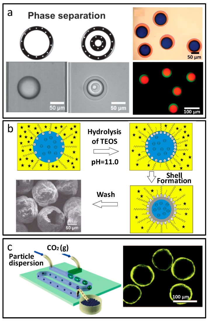
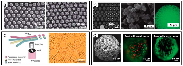
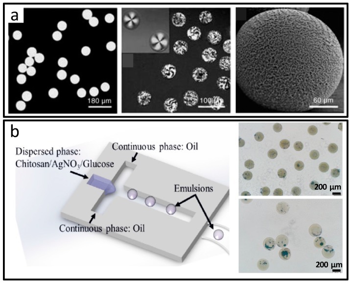
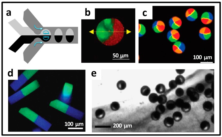
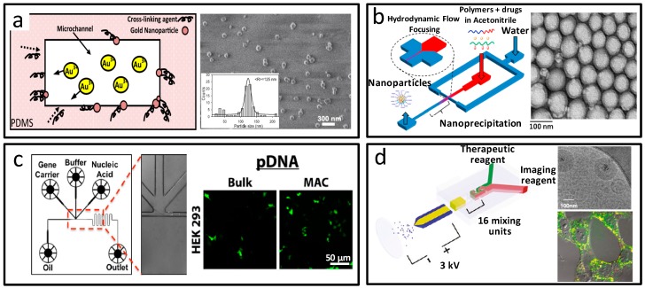
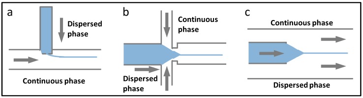
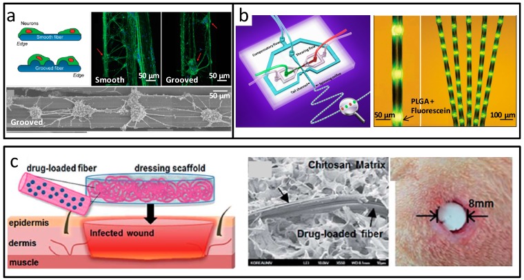
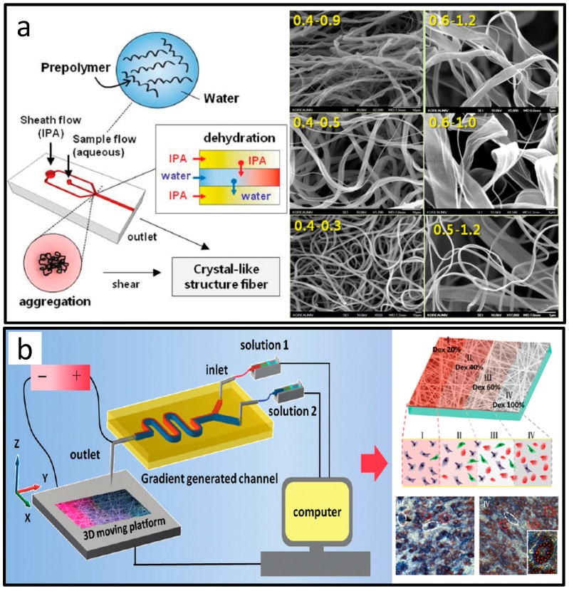
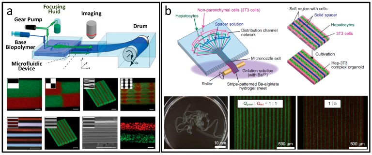
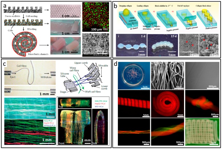
Microfluidics is characterized by laminar flow at micro-scale dimension, high surface to volume ratio, and markedly improved heat/mass transfer. In addition, together with advantages of large-scale integration and flexible manipulation, microfluidic technology has been rapidly developed as one of the most important platforms in the field of functional biomaterial synthesis. Compared to biomaterials assisted by conventional strategies, functional biomaterials synthesized by microfluidics are with superior properties and performances, due to their controllable morphology and composition, which have shown great advantages and potential in the field of biomedicine, biosensing, and tissue engineering. Take the significance of microfluidic engineered biomaterials into consideration; this review highlights the microfluidic synthesis technologies and biomedical applications of materials. We divide microfluidic based biomaterials into four kinds. According to the material dimensionality, it includes: 0D (particulate materials), 1D (fibrous materials), 2D (sheet materials), and 3D (construct forms of materials). In particular, micro/nano-particles and micro/nano-fibers are introduced respectively. This classification standard could include all of the microfluidic biomaterials, and we envision introducing a comprehensive and overall evaluation and presentation of microfluidic based biomaterials and their applications.

Keywords: biological applications; controllable synthesis; functional biomaterials; microfluidics.


Conflict of interest statement

The authors declare no conflict of interest.

Figures

Figure 1
Figure 1
Schematic diagram of the classification of biomaterials engineered from microfluidics.
Figure 2
Figure 2
Microfluidic technologies for the synthesis of particulate materials. The principles and chip designs with different flow regimes for droplet generation, including T-junction (a); flow-focusing (b); and coaxial (c) structured chip; (d) Photolithography applied in microfluidic synthesis.
Figure 3
Figure 3
Spherical microparticles prepared by droplet-based microfluidics. (a) Microspheres from ultraviolet (UV) or heat-induced polymerization in microfluidic droplets; (b) Calcium alginate microbeads for cell encapsulation; (c) Oil-in-water (O/W) droplet synthesis of poly lactic-co-glycolic acid (PLGA) microspheres; (d) SiO2 microspheres generated based on sol-gel principle and microfluidic droplets. Reproduced with permission from [38,40,41,44].
Figure 4
Figure 4
Special-shaped microparticles prepared by droplet-based microfluidics. (a) Poly tripropyleneglycol diacrylate particles with different shapes determined by the microchannel dimension; (b) Polylactic acid (PLA) crescent-shaped composite microparticles from core-shell microcapsules in the microfluidic chip; (c) One-step prepared doughnut-shaped SiO2 microparticles. Reproduced with permission from [45,47,48].
Figure 5
Figure 5
Special-shaped microparticles prepared by photolithography -based microfluidics. Reproduced with permission from [49,50,51].
Figure 6
Figure 6
Core-shell structure microparticles prepared by multiple emulsion-based microfluidics. (a,b) Designs of microfluidic devices for the preparation of double emulsion droplets; (c) Microparticle synthesis with magnetic core and hydrogel shell from double-emulsion microfluidic droplets; (d) Microparticles with two independent oil cores wrapped in the hydrogel shell. Reproduced with permission from [53,54].
Figure 7
Figure 7
Core-shell structure microparticles prepared by phase separation and phase interface effect-based microfluidics. (a) Phase separation process of the polyethylene glycol diacrylate (PEGDA) core-shell microparticles; (b) Hollow SiO2 microspheres from interfacial reaction; (c) Interface self-assembly induced hollow structured microcapsules. Reproduced with permission from [56,58,59].
Figure 8
Figure 8
Porous microparticles prepared by microfluidics. (a) Hierarchically-structured microsphere surface with decorated particles; (b) Microparticles prepared with a network from the surface to the inside from microfluidic O/W droplets; (c) Flow lithography based encoded microparticles; (d) Cell carrier microparticles with controllable pore sizes. Reproduced with permission from [62,63,64,65,67].
Figure 9
Figure 9
Composite microparticles prepared by microfluidics. (a) Microspheres labeled with 4-amino-7-nitrobenzo-2-oxa-1,3-diazole (NBD) dye and CdSe quantum dots; (b) Silver nanoparticle–chitosan composite microparticles assisted by microfluidic synthesis. Reproduced with permission from [45,68].
Figure 10
Figure 10
Janus microparticles prepared by microfluidics. (a) Microfluidic design for Janus droplet generation; (b) Biopolymer-based Janus microbeads with selective degradation; (c) Multi-compartment particles obtained from a multi-barreled capillary; (d) Janus-type, pH-responsive particles via stop-flow lithography; (e) Aggregation and compaction of the Poly(N-isopropylacrylamide) (PNIPAm) microgels. Reproduced with permission from [69,70,71,72].
Figure 11
Figure 11
Nanoparticles prepared by microfluidics. (a) In situ synthesized gold nanoparticles in the microdevices; (b) Dual drug-loaded polymeric nanoparticles by microfluidic self-assembly; (c) Microfluidic preparation of polymer-nucleic acid nanocomplexes; (d) Theranostic lipoplexes synthesized from microfluidic coaxial electrospray. Reproduced with permission from [82,84,89,94].
Figure 12
Figure 12
Microfluidic technologies for the synthesis of fibrous materials. The principles and chip designs with different flow regimes for stable co-flow generation, including T-junction (a); flow-focusing (b); and coaxial (c) structured chip.
Figure 13
Figure 13
Micro-scale fibers prepared by microfluidics. (a) Flat alginate fibers with groove microstructures; (b) Biomimetic bamboo-like hybrid microfibers; (c) Fibrous alginate carrier from microfluidic spinning. Reproduced with permission from [99,103,105].
Figure 14
Figure 14
Nano-scale fibers prepared by microfluidics. (a) Micro/nanometer-scale fiber with ordered structures; (b) Gradient electrospinning nanofibers assisted by microfluidics. Reproduced with permission from [108,109].
Figure 15
Figure 15
Sheet materials prepared by microfluidics. (a) Mosaic hydrogels sheets, scale bars are all 500 μm; (b) Micronozzle device patterned hydrogel sheet. Reproduced with permission from [110,112].
Figure 16
Figure 16
Construct forms of materials prepared by microfluidics. (a) Bottom-up fabrication of 3D microfluidic cell-laden constructs; (b) Collagen building constructs for self-assembly of 3D microtissues; (c) Microfiber-based assembly of 3D macroscopic cellular structures; (d) Constructs made by the automated assembly of in situ formed microfibers. Reproduced with permission from [115,116,120,122].
Figure 17
Figure 17
Scaled-up synthesis of functional biomaterials based on microfluidics. (a) High-volume production of emulsions in a microfluidic parallelization arrangement; (b) A liter per hour volume production of single emulsions in a parallelization chip; (c) Particle synthesis in parallel multiple modular microfluidic reactors. Reproduced with permission from [123,124,125].

References

    1. Ríos Á., Zougagh M., Avila M. Miniaturization through lab-on-a-chip: Utopia or reality for routine laboratories? A review. Anal. Chim. Acta. 2012;740:1–11. doi: 10.1016/j.aca.2012.06.024. - DOI - PubMed
    1. Kricka L.J. Miniaturization of analytical systems. Clin. Chem. 1998;44:2008–2014. - PubMed
    1. Sauer S., Lange B.M.H., Gobom J., Nyarsik L., Seitz H., Lehrach H. Miniaturization in functional genomics and proteomics. Nat. Rev. Genet. 2005;6:465–476. doi: 10.1038/nrg1618. - DOI - PubMed
    1. Whitesides G.M. The origins and the future of microfluidics. Nature. 2006;442:368–373. doi: 10.1038/nature05058. - DOI - PubMed
    1. Dittrich P.S., Manz A. Lab-on-a-chip: Microfluidics in drug discovery. Nat. Rev. Drug Discov. 2006;5:210–218. doi: 10.1038/nrd1985. - DOI - PubMed

LinkOut - more resources