. 2018 Nov 6;16(1):302.
doi: 10.1186/s12967-018-1676-3.

Periostin involved in the activated hepatic stellate cells-induced progression of residual hepatocellular carcinoma after sublethal heat treatment: its role and potential for therapeutic inhibition

Affiliations

Periostin involved in the activated hepatic stellate cells-induced progression of residual hepatocellular carcinoma after sublethal heat treatment: its role and potential for therapeutic inhibition

Rui Zhang et al. J Transl Med. .

Abstract

Background: Incomplete thermal ablation may induce invasiveness of hepatocellular carcinoma (HCC). Here, we investigated whether activated hepatic stellate cells (HSCs) would accelerate the progression of residual HCC after sublethal heat treatment, and thus sought to identify the potential targets.

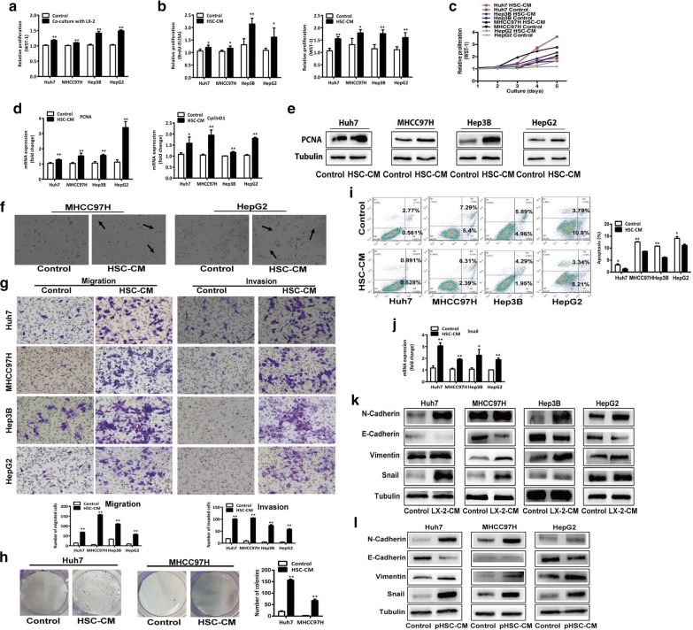
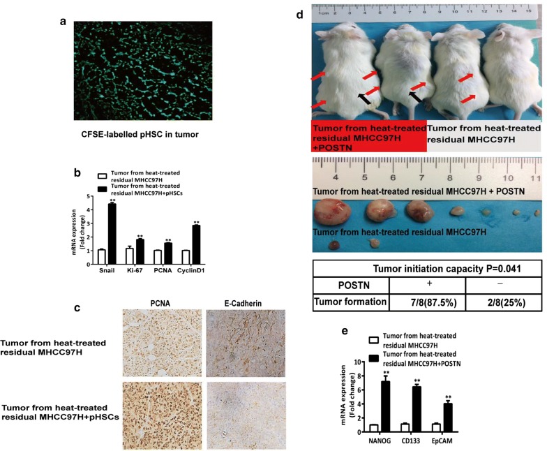
Methods: Hepatocellular carcinoma cells were exposed to sublethal heat treatment and then cultured with the conditioned medium from activated HSCs (HSC-CM). The cell proliferation, migration, invasion and parameters of epithelial-mesenchymal transition (EMT) were analyzed. In vivo tumor progression of heat-treated residual HCC cells inoculated with activated HSCs was studied in nude mice.

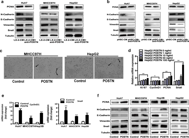
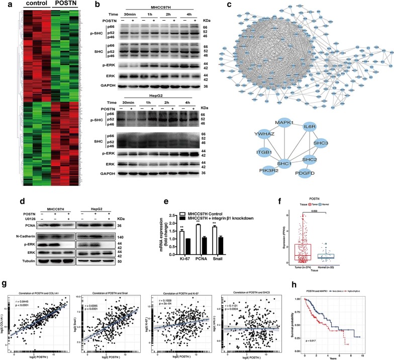
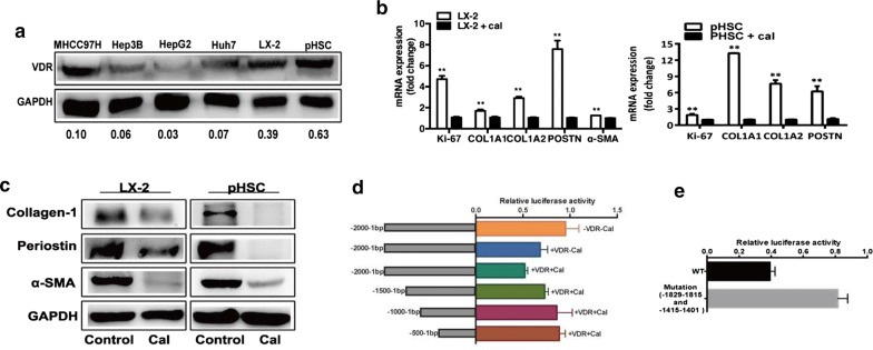
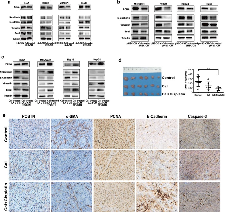
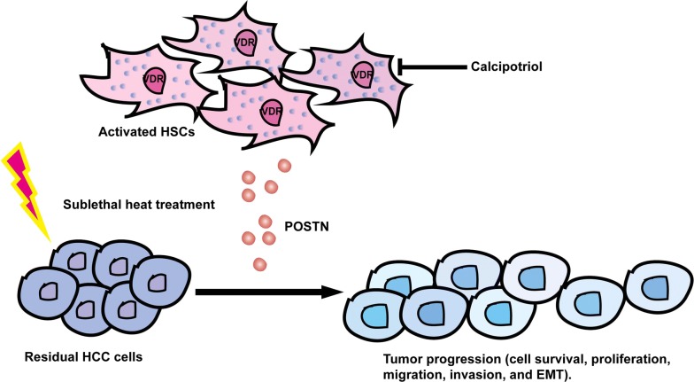
Results: HSC-CM significantly enhanced the proliferation, motility, invasion, prominent EMT activation and decreased apoptosis of heat-exposed residual HCC cells. These increased malignant phenotypes were markedly attenuated by neutralizing periostin (POSTN) in HSC-CM. Furthermore, exogenous POSTN administration exerted the similar effects of HSC-CM on heat-treated residual HCC cells. POSTN induced the prominent activation of p52Shc and ERK1/2 via integrin β1 in heat-exposed residual HCC cells. Vitamin D analog calcipotriol blocked POSTN secretion from activated HSCs. Calcipotriol plus cisplatin significantly suppressed the activated HSCs-enhanced tumor progression of heat-treated residual HCC cells via the inhibited POSTN expression and the increased apoptosis.

Conclusions: Activated HSCs promote the tumor progression of heat-treated residual HCC through the release of POSTN, which could be inhibited by calcipotriol. Calcipotriol plus cisplatin could be used to thwart the accelerated progression of residual HCC after suboptimal heat treatment.

Keywords: Calcipotriol; Hepatic stellate cells; Hepatocellular carcinoma; Periostin.


Figures

Fig. 1
Fig. 1
Activated HSCs or HSC-CM promoted the proliferation, migration, invasion, colony formation, and decreased the apoptosis of heat-exposed residual HCC cells. a When heat-treated residual HCC cells (Huh7, MHCC97H, Hep3B and HepG2) were co-cultured with activated HSCs LX2, proliferation was significantly higher than the cells cultured alone, as analyzed by WST-1 assay. b, c Proliferation of heat-treated residual HCC cells cultured in HSC-CM from LX2 cells was significantly higher than the cells cultured with control medium, as indicated by the WST-1 assay, BrdU-ELISA and growth curves. d PCNA and cyclinD1 mRNA expression of heat-treated residual HCC cells cultured in HSC-CM from LX2 cells were significantly higher than the cells cultured with control medium, as measured by qRT-PCR. e PCNA protein expression was analyzed by western blot. f Heat-treated residual HCC cells displayed distinct spindle-like appearance when cultured with HSC-CM from LX2 cells. g HSC-CM from LX2 cells promoted the in vitro migration and invasion of heat-treated residual HCC cells. The number of migrated and invaded cells was counted. h Colony formation of heat-treated residual HCC cells cultured in HSC-CM from LX2 cells was increased. i Apoptosis of heat-treated residual HCC cells in the presence of HSC-CM from LX2 cells was reduced than the cell cultured with control medium, as analyzed by Annexin V/PI staining. jl The levels of Snail mRNA expression, and Snail, Vimentin, E-cadherin, and N-cadherin protein expression in heat-treated residual HCC cells cultured with HSC-CM from LX2 cells or pHSC cells compared with control medium were analyzed by qRT-PCR and western blot. pHSC primary hepatic stellate cells. **P < 0.01; *P < 0.05
Fig. 2
Fig. 2
POSTN in HSC-CM mediated the increased proliferation, migration, and invasion of heat-exposed residual HCC cells. a, b The alterations of Snail, Vimentin, E-cadherin, N-cadherin, PCNA protein expression in heat-exposed residual HCC cells cultured with POSTN-depleting HSC-CM from LX2 cells or pHSC cells. c Distinct spindle-like appearance of heat-treated residual HCC cells (MHCC97H and HepG2) induced by exogenous POSTN (100 ng/mL). d After treated with exogenous POSTN, heat-exposed residual HepG2 cells showed significantly higher levels of Ki-67, cyclinD1, PCNA, Snail mRNA expression in a dose-dependent manner, as measured by qRT-PCR. e, f After treated with exogenous POSTN, heat-exposed residual HCC cells (Huh7, MHCC97H, HepG2 and Hep3B) showed significantly higher levels of cyclinD1, Snail, PCNA, N-cadherin, Vimentin and markedly lower level of E-cadherin compared to the control, as detected by qRT-PCR and western blot. pHSC primary hepatic stellate cells. **P < 0.01; *P < 0.05
Fig. 3
Fig. 3
POSTN induced the Shc-ERK activation of heat-exposed residual HCC cells through integrin β1. a The mRNA expression profile of heat-treated residual MHCC97H cells in response to POSTN was illustrated as a heatmap. Red, green represent high and low mRNA expression. b With POSTN treatment, the phosphorylated of p52Shc and ERK1/2 in heat-exposed residual HCC cells (MCHCC97H and HepG2) were significantly increased in a time-dependent manner. c PPI network analysis of the differentially expressed genes identified Shc as a gene of biological importance in POSTN-mediated signaling networks and a diagram illustrated the interaction of Shc with the molecules (e.g., ITGB1 and MAPK1). d When heat-exposed residual HCC cells (MCHCC97H and HepG2) were treated with POSTN, the levels of PCNA, N-cadherin and ERK1/2 phosphorylation were significantly increased. ERK1/2 inhibitor (U0126, 25 μM) reversed the above POSTN-induced increase. e With the stimulation of exogenous POSTN, the levels of Ki-67, PCNA and Snail mRNA expression were significantly decreased in heat-exposed residual integrin β1-knockdown MHCC97H cells. f Expression of POSTN in HCC tissues (n = 374) than that of adjacent non-tumor tissues (n = 50) in the HCC data of TCGA cohorts. g A significant positive correlation between the degree of POSTN expression also showed with that of COL1A1 (r = 0.8445, P < 0.0001), Ki-67 (r = 0.1928, P = 2×10⁻⁴), Snail (r = 0.6395, P < 0.0001), and Sch3 (r = 0.1121, P = 0.0304) in the TCGA-HCC cohorts. h HCC patients were stratified by POSTN and MAPK1 (ERK2) expression and the co-expression of POSTN and ERK2 predicted poor-survival prognosis in the TCGA-HCC cohorts by Kaplan–Meier analyses
Fig. 4
Fig. 4
Activated HSCs promoted the in vivo tumor progression of heat-treated residual HCC cells. a HSCs cells were found in the tumors at 2 weeks after the inoculation of heat-treated residual MHCC97H cells with CFSE-labelled pHSCs. b, c Up-regulation of Ki-67, PCNA, cyclin D1 and Snail mRNA expression was found in the tumors formed from heat-exposed residual MHCC97H and pHSCs compared with the tumors from heat-exposed residual MHCC97H alone, as detected by qRT-PCR. The protein expression of PCNA and E-Cadherin was shown by immunohistochemical analysis. d Xenograft tumorigenicity assay. Heat-exposed residual MHCC97H (2 × 104 cells) with 100 ng/mL POSTN subcutaneously inoculated into NOD/SCID mice developed more and larger tumors compared to the cells injected alone. Red arrows on the left flank represent the tumors formed by heat-exposed residual MHCC97H cells with 100 ng/mL POSTN while black arrows on the right flank represent the tumors formed by heat-exposed residual MHCC97H cells alone. The number and images of tumors developed in mice after 8 weeks are shown. The two groups were compared by using Fisher exact test. e The mRNA expression of NANOG, CD133, EpCAM was significantly up-regulated in the tumors generated from heat-exposed residual MHCC97H cells with 100 ng/mL POSTN. **P < 0.01; *P < 0.05
Fig. 5
Fig. 5
VDR regulates the expression of POSTN in presence of calcipotriol. a Vitamin D receptor (VDR) expression in activated HSCs (LX2 and pHSC) and HCC cell lines (MHCC97H, Hep3B, HepG2 and Huh7) was detected by Western blot. b The markers and activities of activated HSCs (α-SMA, COL1A1, COL1A2, Ki-67, and POSTN) were down-regulated in LX2 cells and pHSC cells by calcipotriol treatment, as analyzed using qRT-PCR. c Protein expression of α-SMA, POSTN and collagen-1 were reduced in LX2 cells and pHSC cells by calcipotriol treatment, as measured by western blot. d Results of Luciferase reporter assay for POSTN promoter constructs. A ~ 2-kb-long POSTN promoter and its serial deletion constructs were measured in HEK 293T cells and results of the firefly luciferase were normalized to the Renilla luciferase activity. e Luciferase activity of mutant reporter plasmids that the two putative VDR transcription factor binding sites were altered by site-directed mutagenesis in the full-length POSTN promoter. Calcipotriol, Cal. VDR, VDR-expressing plasmid. **P < 0.01; *P < 0.05
Fig. 6
Fig. 6
Calcipotriol suppressed the activated HSCs-induced tumor progression of heat-treated residual HCC cells in vivo. a, b The changes of Snail, Vimentin, E-cadherin and N-cadherin, PCNA expression in heat-exposed residual HCC cells (MHCC97H, Hep3B, HepG2 and Huh7) cultured with conditioned medium (CM) from HSCs or calcipotriol-treated HSCs were compared. HSCs cells (LX2 and pHSC) were stimulated by the pre-treatment with TGF-β1. c The changes of Snail, Vimentin, E-cadherin and N-cadherin, PCNA expression in heat-treated residual HCC cells were reversed when CM from calcipotriol-treated LX2 cells was supplemented with 100 ng/mL POSTN. d Calcipotriol inhibited tumor growth compared with the control group. The combination therapy of calcipotriol and cisplatin significantly reduced tumor growth. e The expression of POSTN, α-SMA, PCNA, E-Cadherin and cleaved-Caspase-3 was detected by immunohistochemical analysis. Calcipotriol, Cal. **P < 0.01; *P < 0.05
Fig. 7
Fig. 7
A proposed graphical diagram that activated HSCs promote tumor progression of heat-treated residual HCC cells via POSTN secretion which could be inhibited by calcipotriol

Similar articles

Cited by

References

    1. Shiina S, Tateishi R, Arano T, Uchino K, Enooku K, Nakagawa H, et al. Radiofrequency ablation for hepatocellular carcinoma: 10-year outcome and prognostic factors. Am J Gastroenterol. 2012;107:569–577. doi: 10.1038/ajg.2011.425. - DOI - PMC - PubMed
    1. Lee DH, Lee JM, Lee JY, Kim SH, Yoon JH, Kim YJ, et al. Radiofrequency ablation of hepatocellular carcinoma as first-line treatment: long-term results and prognostic factors in 162 patients with cirrhosis. Radiology. 2014;270:900–909. doi: 10.1148/radiol.13130940. - DOI - PubMed
    1. Yin XY, Xie XY, Lu MD, Xu HX, Xu ZF, Kuang M, et al. Percutaneous thermal ablation of medium and large hepatocellular carcinoma: long-term outcome and prognostic factors. Cancer. 2009;115:1914–1923. doi: 10.1002/cncr.24196. - DOI - PubMed
    1. Seror O, N’Kontchou G, Ibraheem M, Ajavon Y, Barrucand C, Ganne N, et al. Large (≥ 5.0-cm) HCCs: multipolar RF ablation with three internally cooled bipolar electrodes—initial experience in 26 patients. Radiology. 2008;248:288–296. doi: 10.1148/radiol.2481071101. - DOI - PubMed
    1. Livraghi T, Goldberg SN, Lazzaroni S, Meloni F, Ierace T, Solbiati L, et al. Hepatocellular carcinoma: radio-frequency ablation of medium and large lesions. Radiology. 2000;214:761–768. doi: 10.1148/radiology.214.3.r00mr02761. - DOI - PubMed

Publication types

MeSH terms