

Foetal weight prediction models at a given gestational age in the absence of ultrasound facilities: application in Indonesia
- PMID: 30400880
- PMCID: PMC6219176
- DOI: 10.1186/s12884-018-2047-z
Foetal weight prediction models at a given gestational age in the absence of ultrasound facilities: application in Indonesia
Abstract
Background: Birth weight is one of the most important indicators of neonatal survival. A reliable estimate of foetal weight at different stages of pregnancy would facilitate intervention plans for medical practitioners to prevent the risk of low birth weight delivery. This study has developed reliable models to more accurately predict estimated foetal weight at a given gestation age in the absence of ultrasound facilities.
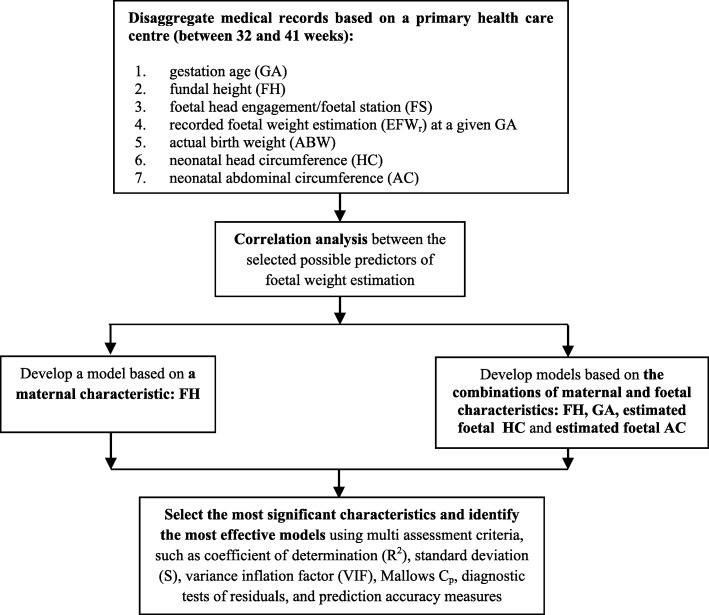
Methods: A primary health care centre was involved in collecting retrospective non-identified Indonesian data. The best subset model selection criteria, coefficient of determination, standard deviation, variance inflation factor, Mallows Cp, and diagnostic tests of residuals were deployed to select the most significant independent variables. Simple and multivariate linear regressions were used to develop the proposed models. The efficacy of models for predicting foetal weight at a given gestational age was assessed using multi-prediction accuracy measures.
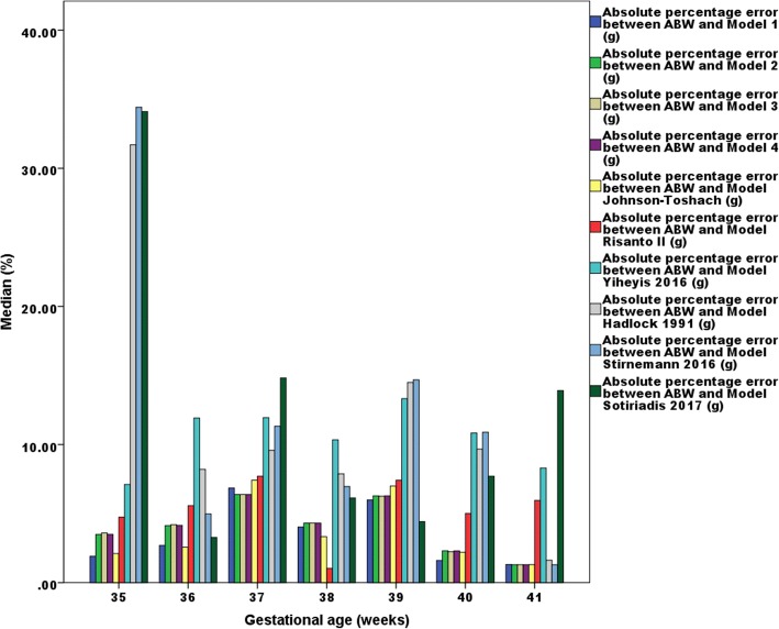
Results: Four weight prediction models based on fundal height and its combinations with gestational age (between 32 and 41 weeks) and ultrasonic estimates of foetal head circumference and foetal abdominal circumference have been developed. Multiple comparison criteria show that the proposed models were more accurate than the existing models (mean prediction errors between - 0.2 and 2.4 g and median absolute percentage errors between 4.1 and 4.2%) in predicting foetal weight at a given gestational age (between 35 and 41 weeks).
Conclusions: This research has developed models to more accurately predict estimated foetal weight at a given gestational age in the absence of ultrasound machines and trained ultra-sonographers. The efficacy of the models was assessed using retrospective data. The results show that the proposed models produced less error than the existing clinical and ultrasonic models. This research has resulted in the development of models where ultrasound facilities do not exist, to predict the estimated foetal weight at varying gestational age. This would promote the development of foetal inter growth charts, which are currently unavailable in Indonesian primary health care systems. Consistent monitoring of foetal growth would alleviate the risk of having inter growth abnormalities, such as low birth weight that is the most leading factor of neonatal mortality.
Keywords: Absence of ultrasound facilities; Estimated foetal abdominal circumference; Estimated foetal head circumference; Foetal weight estimation; Fundal height; Gestational age; Indonesia; Prediction accuracy; Primary health care centre; Regression analysis.
Conflict of interest statement
Authors’ information
DA: PhD candidate in the Mathematical Sciences (Applied Statistics), School of Science (Mathematical and Geospatial Sciences), College of Science, Engineering, and Health, RMIT University, Melbourne, Australia and Junior Lecturer at Study Program of Statistics, Faculty of Mathematics and Natural Sciences, University of Lambung Mangkurat (ULM), South Kalimantan, Indonesia.
MA: Senior Lecturer of Statistical Quality Control and its applications in: manufacturing industry, air pollution control, software quality, univariate and multivariate processes, health industry, and the banking system, School of Science (Mathematical and Geospatial Sciences), College of Science, Engineering, and Health, RMIT University, Melbourne, Australia.
KM: Senior Lecturer of Applied Statistics and Mathematics, Market Research, and Numerical Analysis in aerospace engineering, clinical sciences, geomatic engineering, and oncology and carcinogenesis, College of Science, Engineering, and Health, RMIT University, Melbourne, Australia.
Ethics approval and consent to participate
This study is a part of doctoral degree research and has obtained two ethics clearances:
The Ethical Committees of Medical Research, Medical Faculty, University of Lambung Mangkurat (ULM), Banjarmasin, South Kalimantan (Indonesia), on March 10th, 2016, with registration number: 018/KEPK-FK UNLAM/EC/III/2016. Permission to access unidentified secondary data in the preganancy register available at the selected primary health care was also granted under this ethical consideration.
The Science, Engineering, and Health College Human Ethics Advisory Network (CHEAN) of Royal Melbourne Institute of Technology (RMIT) University, Melbourne, Victoria (Australia), on March 16th, 2016, with registration number: ASEHAPP 19-16/RM No: 19974.
Research permissions were obtained from the Indonesian national, provincial, and local governments.
Consent for publication
The manuscript does not contain any individual person’s data; hence consent for publication is not applicable.
Competing interests
The authors declare that they have no competing interests.
Publisher’s Note
Springer Nature remains neutral with regard to jurisdictional claims in published maps and institutional affiliations.
Figures

Similar articles
-
Prediction of birthweight and risk of macrosomia in pregnancies complicated by diabetes.Am J Obstet Gynecol MFM. 2023 Aug;5(8):101042. doi: 10.1016/j.ajogmf.2023.101042. Epub 2023 Jun 6. Am J Obstet Gynecol MFM. 2023. PMID: 37286100
-
Two-stage approach for prediction of small-for-gestational-age neonate and adverse perinatal outcome by routine ultrasound examination at 35-37 weeks' gestation.Ultrasound Obstet Gynecol. 2019 Oct;54(4):484-491. doi: 10.1002/uog.20391. Epub 2019 Aug 27. Ultrasound Obstet Gynecol. 2019. PMID: 31271475
-
Comparison of fundal height measurement and sonographically measured fetal abdominal circumference in the prediction of high and low birth weight at term.Ultrasound Obstet Gynecol. 2009 Nov;34(5):566-71. doi: 10.1002/uog.6378. Ultrasound Obstet Gynecol. 2009. PMID: 19582801
-
Diagnostic performance of third-trimester ultrasound for the prediction of late-onset fetal growth restriction: a systematic review and meta-analysis.Am J Obstet Gynecol. 2019 May;220(5):449-459.e19. doi: 10.1016/j.ajog.2018.09.043. Epub 2019 Jan 8. Am J Obstet Gynecol. 2019. PMID: 30633918
-
Magnetic resonance imaging for prenatal estimation of birthweight in pregnancy: review of available data, techniques, and future perspectives.Am J Obstet Gynecol. 2019 May;220(5):428-439. doi: 10.1016/j.ajog.2018.12.031. Epub 2018 Dec 22. Am J Obstet Gynecol. 2019. PMID: 30582928 Review.
Cited by
-
Development and validation of prediction models for fetal growth restriction and birthweight: an individual participant data meta-analysis.Health Technol Assess. 2024 Aug;28(47):1-119. doi: 10.3310/DABW4814. Health Technol Assess. 2024. PMID: 39252507 Free PMC article.
-
The Impact of Scientific and Technical Training on Improving Databases' Adequacy for Fetal Growth Chart Development in Limited-Resource Settings: A Case Study in the Province of South Kalimantan, Indonesia.J Pregnancy. 2019 Feb 3;2019:8540637. doi: 10.1155/2019/8540637. eCollection 2019. J Pregnancy. 2019. PMID: 30854237 Free PMC article.
-
The development of an alternative growth chart for estimated fetal weight in the absence of ultrasound: Application in Indonesia.PLoS One. 2020 Oct 13;15(10):e0240436. doi: 10.1371/journal.pone.0240436. eCollection 2020. PLoS One. 2020. PMID: 33048951 Free PMC article. Clinical Trial.
-
Improving the Information Availability and Accessibility of Antenatal Measurements to Ensure Safe Delivery: A Research-Based Policy Recommendation to Reduce Neonatal Mortality in Indonesia.Int J Womens Health. 2020 May 6;12:369-380. doi: 10.2147/IJWH.S247213. eCollection 2020. Int J Womens Health. 2020. PMID: 32440231 Free PMC article.
-
Estimation of Gestational Age Using Neonatal Anatomical Anthropometric Parameters in Dessie Referral Hospital, Northeast Ethiopia.Risk Manag Healthc Policy. 2020 Dec 15;13:3021-3029. doi: 10.2147/RMHP.S280682. eCollection 2020. Risk Manag Healthc Policy. 2020. PMID: 33376426 Free PMC article.
References
-
- Parvin Z, Shafiuddin S, Uddin MA, Begum F. Symphysio fundal height (SFH) measurement as a predictor of birth weight. Faridpur Med Coll J. 2013;7(2):54–58. doi: 10.3329/fmcj.v7i2.13498. - DOI
MeSH terms
LinkOut - more resources
Full Text Sources
Medical
Molecular Biology Databases
Miscellaneous
