The myocardial infarct-exacerbating effect of cell-free DNA is mediated by the high-mobility group box 1-receptor for advanced glycation end products-Toll-like receptor 9 pathway
- PMID: 30401529
- PMCID: PMC6450791
- DOI: 10.1016/j.jtcvs.2018.09.043
The myocardial infarct-exacerbating effect of cell-free DNA is mediated by the high-mobility group box 1-receptor for advanced glycation end products-Toll-like receptor 9 pathway
Abstract
Introduction: Damage-associated molecular patterns, such as high-mobility group box 1 (HMGB1) and cell-free DNA (cfDNA), play critical roles in mediating ischemia-reperfusion injury (IRI). HMGB1 activates RAGE to exacerbate IRI, but the mechanism underlying cfDNA-induced myocardial IRI remains unknown. We hypothesized that the infarct-exacerbating effect of cfDNA is mediated by HMGB1 and receptor for advanced glycation end products (RAGE).
Methods: C57BL/6 wild type mice, RAGE knockout (KO), and Toll-like receptor 9 KO mice underwent 20- or 40-minute occlusions of the left coronary artery followed by up to 60 minutes of reperfusion. Cardiac coronary perfusate was acquired from ischemic hearts without reperfusion. Exogenous mitochondrial DNA was acquired from livers of normal C57BL/6 mice. Myocardial infarct size (IS) was reported as percent risk region, as measured by 2,3,5-triphenyltetrazolium chloride and Phthalo blue (Heucotech, Fairless Hill, Pa) staining. cfDNA levels were measured by Sytox Green assay (Thermo Fisher Scientific, Waltham, Mass) and/or spectrophotometer.
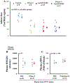
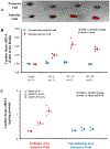
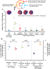
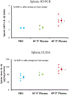
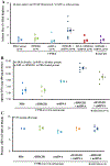
Results: Free HMGB1 and cfDNA levels were increased in the ischemic myocardium during prolonged ischemia and subsequently in the plasma during reperfusion. In C57BL/6 mice undergoing 40'/60' IRI, deoxyribonuclease I, or anti-HMGB1 monoclonal antibody reduced IS by approximately half to 29.0% ± 5.2% and 24.3% ± 3.5% (P < .05 vs control 54.3% ± 3.4%). However, combined treatment with deoxyribonuclease I + anti-HMGB1 monoclonal antibody did not further attenuate IS (29.3% ± 4.9%). In C57BL/6 mice undergoing 20'/60' IRI, injection of 40'/5' plasma upon reperfusion increased IS by more than 3-fold (to 19.9 ± 4.3; P < .05). This IS exacerbation was abolished by pretreating the plasma with deoxyribonuclease I or by depleting the HMGB1 by immunoprecipitation, or by splenectomy. The infarct-exacerbating effect also disappeared in RAGE KO mice and Toll-like receptor 9 KO mice. Injection of 40'/0' coronary perfusate upon reperfusion similarly increased IS. The levels of HMGB1 and cfDNA were significantly elevated in the 40'/0' coronary perfusate and 40'/reperfusion (min) plasma but not in those with 10' ischemia. In C57BL/6 mice without IRI, 40'/5' plasma significantly increased the interleukin-1β protein and messenger RNA expression in the spleen by 30 minutes after injection. Intravenous bolus injection of recombinant HMGB1 (0.1 μg/g) or mitochondrial DNA (0.5 μg/g) 5 minutes before reperfusion did not exacerbate IS (P = not significant vs control). However, combined administration of recombinant HMGB1 + mitochondrial DNA significantly increased IS (P < .05 vs individual treated groups) and this infarct-exacerbating effect disappeared in RAGE KO mice and splenectomized C57BL/6 mice. The accumulation of cfDNA in the spleen after combined recombinant HMGB1 + mitochondrial DNA treatment was significantly more elevated in C57BL/6 mice than in RAGE KO mice.
Conclusions: Both HMGB1 and cfDNA are released from the heart upon reperfusion after prolonged ischemia and both contribute importantly and interdependently to post-IRI by a common RAGE-Toll-like receptor 9-dependent mechanism. Depleting either of these 2 damage-associated molecular patterns suffices to significantly reduce IS by approximately 50%.
Keywords: HMGB1; RAGE; TLR9; cell-free DNA; ischemia/reperfusion; spleen.
Copyright © 2018 The American Association for Thoracic Surgery. Published by Elsevier Inc. All rights reserved.
Figures



Comment in
-
Commentary: RAGE against the spleen.J Thorac Cardiovasc Surg. 2019 Jun;157(6):2272-2273. doi: 10.1016/j.jtcvs.2018.09.071. Epub 2018 Oct 9. J Thorac Cardiovasc Surg. 2019. PMID: 30385018 No abstract available.
-
Commentary: Circulating factors released after myocardial infarction: Beneficial or detrimental?J Thorac Cardiovasc Surg. 2019 Jun;157(6):2270-2271. doi: 10.1016/j.jtcvs.2018.10.128. Epub 2018 Nov 8. J Thorac Cardiovasc Surg. 2019. PMID: 30503734 No abstract available.
References
-
- Liu J, Cai X, Xie L, et al. Circulating Cell Free Mitochondrial DNA is a Biomarker in the Development of Coronary Heart Disease in the Patients with Type 2 Diabetes. Clin Lab. 2015;61:661–667. - PubMed
-
- Xie L, Liu S, Cheng J, Wang L, Liu J, Gong J. Exogenous administration of mitochondrial DNA promotes ischemia reperfusion injury via TLR9-p38 MAPK pathway. Regul Toxicol Pharmacol. 2017;89:148–154. - PubMed
MeSH terms
Substances
Grants and funding
LinkOut - more resources
Full Text Sources
Medical
Research Materials

