Review
. 2019 Jan 3;133(1):59-69.
doi: 10.1182/blood-2018-08-815951. Epub 2018 Nov 6.

The molecular genetics of sideroblastic anemia

Affiliations
Review

The molecular genetics of sideroblastic anemia

Sarah Ducamp et al. Blood. .

Abstract

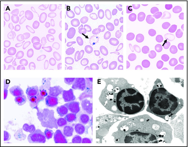
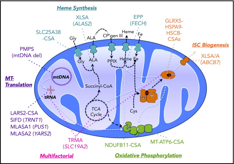
The sideroblastic anemias (SAs) are a group of inherited and acquired bone marrow disorders defined by pathological iron accumulation in the mitochondria of erythroid precursors. Like most hematological diseases, the molecular genetic basis of the SAs has ridden the wave of technology advancement. Within the last 30 years, with the advent of positional cloning, the human genome project, solid-state genotyping technologies, and next-generation sequencing have evolved to the point where more than two-thirds of congenital SA cases, and an even greater proportion of cases of acquired clonal disease, can be attributed to mutations in a specific gene or genes. This review focuses on an analysis of the genetics of these diseases and how understanding these defects may contribute to the design and implementation of rational therapies.


Conflict of interest statement

Conflict-of-interest disclosure: The authors declare no competing financial interests.

Figures

Figure 1.
Figure 1.
Morphological features of SA. (A) May-Gruenwald-Giemsa (MGG)-stained peripheral blood smear of a man with mild XLSA demonstrating hypochromia, anisocytosis, and microcytosis. (B) Iron-stained peripheral blood smear from the same patient highlighting a siderocyte (arrow). (C) MGG-stained peripheral blood smear from the patient’s mother demonstrating the dimorphic red blood cell population, including hypochromic microcytes containing Pappenheimer bodies (arrow). (D) Iron-stained bone marrow aspirate smear from a man with XLSA demonstrating iron granules (blue) ringing around late erythroblast nuclei. (E) Transmission electron micrograph from a patient with RARS demonstrating electron densities (black) within degenerating mitochondria (pale vacuoles, indicated by an arrow) ringing around erythroblast nuclei (photograph courtesy of Marcel Seiler, Boston Veterans Affairs Medical Center [VAMC], Boston, MA). (A-D) Equally scaled; original magnification ×1000. (E) Original magnification ∼×8000.
Figure 2.
Figure 2.
Pathways and genes implicated in CSA. Mitochondrial pathways relevant to the pathogenesis of CSA are diagrammed. Synthetic pathways are indicated by black dashed lines, whereas pathways whose products are required for other pathways implicated in CSA are shown in gray lines. Heme biosynthetic, iron-sulfur cluster biogenesis, mitochondrial (MT) translation, and oxidative phosphorylation/mitochondrial respiration pathways and the genes mutated in CSA in those pathways are highlighted in distinct colors. Thiamine-responsive megaloblastic anemia (TRMA) affects multiple pathways dependent upon thiamine. CP, coproporphyrin; Gly, glycine; MLASA, mitochondrial myopathy with lactic acidosis and SA; mtDNA, mitocondrial DNA; PMPS, Pearson marrow-pancreas syndrome; PPIX, protoporphyrin IX; TCA, tricarboxylic acid. Modified from Fleming with permission.

References

    1. Cartwright GE, Deiss A. Sideroblasts, siderocytes, and sideroblastic anemia. N Engl J Med. 1975;292(4):185-193. - PubMed
    1. Fleming MD. The genetics of inherited sideroblastic anemias. Semin Hematol. 2002;39(4):270-281. - PubMed
    1. Bottomley SS, Fleming MD. Sideroblastic anemia: diagnosis and management. Hematol Oncol Clin North Am. 2014;28(4):653-670. - PubMed
    1. Dacie JV, Doniach I. The basophilic property of the iron-containing granules in siderocytes. J Pathol. 1947;59(4):684-686. - PubMed
    1. McFadzean AJ, Davis LJ. Iron-staining erythrocytic inclusions with especial reference to acquired haemolytic anaemia. Glasg Med J. 1947;28(9):237-279. - PMC - PubMed

Publication types

MeSH terms