. 2018 Nov 6;11(555):eaaq1075.
doi: 10.1126/scisignal.aaq1075.

Biased agonists of the chemokine receptor CXCR3 differentially control chemotaxis and inflammation

Affiliations

Biased agonists of the chemokine receptor CXCR3 differentially control chemotaxis and inflammation

Jeffrey S Smith et al. Sci Signal. .

Abstract

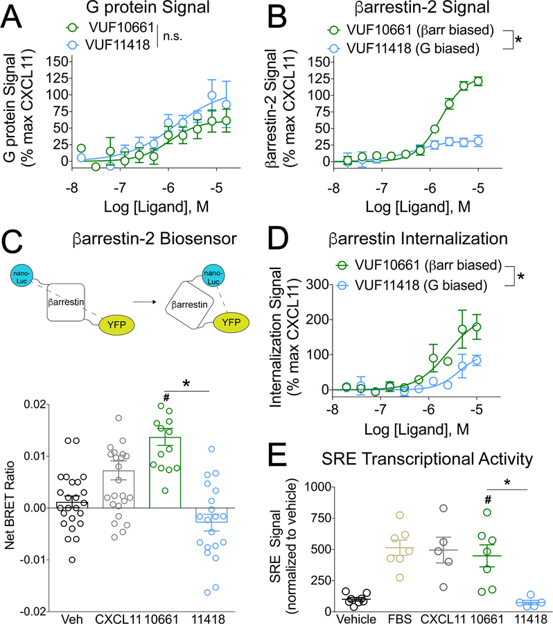
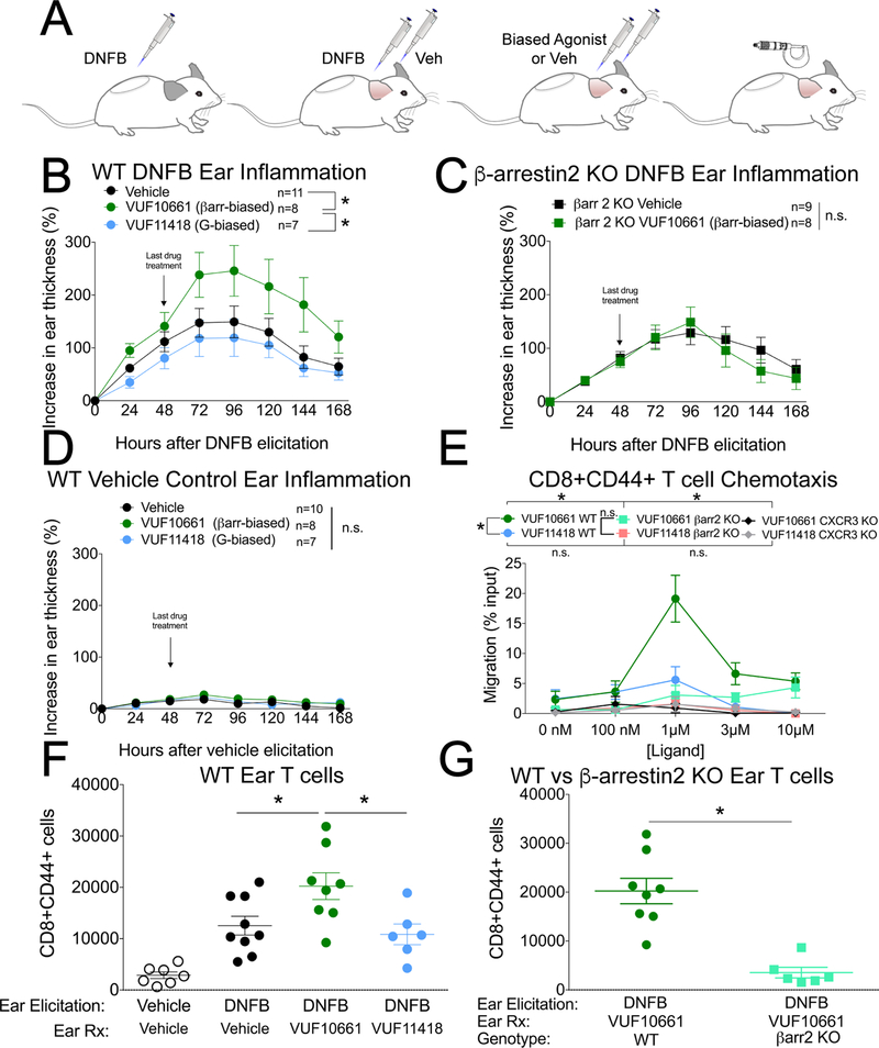
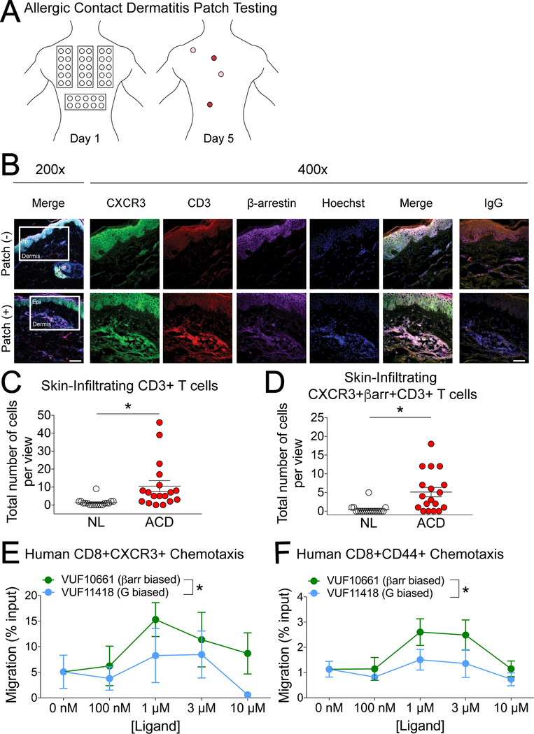
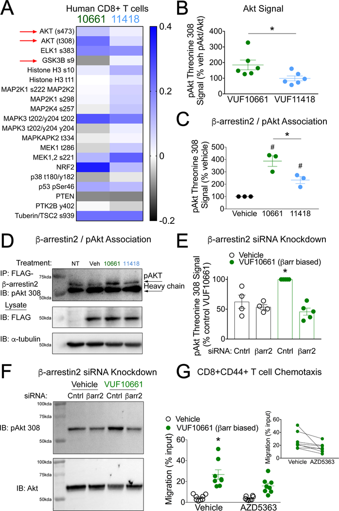
The chemokine receptor CXCR3 plays a central role in inflammation by mediating effector/memory T cell migration in various diseases; however, drugs targeting CXCR3 and other chemokine receptors are largely ineffective in treating inflammation. Chemokines, the endogenous peptide ligands of chemokine receptors, can exhibit so-called biased agonism by selectively activating either G protein- or β-arrestin-mediated signaling after receptor binding. Biased agonists might be used as more targeted therapeutics to differentially regulate physiological responses, such as immune cell migration. To test whether CXCR3-mediated physiological responses could be segregated by G protein- and β-arrestin-mediated signaling, we identified and characterized small-molecule biased agonists of the receptor. In a mouse model of T cell-mediated allergic contact hypersensitivity (CHS), topical application of a β-arrestin-biased, but not a G protein-biased, agonist potentiated inflammation. T cell recruitment was increased by the β-arrestin-biased agonist, and biopsies of patients with allergic CHS demonstrated coexpression of CXCR3 and β-arrestin in T cells. In mouse and human T cells, the β-arrestin-biased agonist was the most efficient at stimulating chemotaxis. Analysis of phosphorylated proteins in human lymphocytes showed that β-arrestin-biased signaling activated the kinase Akt, which promoted T cell migration. This study demonstrates that biased agonists of CXCR3 produce distinct physiological effects, suggesting discrete roles for different endogenous CXCR3 ligands and providing evidence that biased signaling can affect the clinical utility of drugs targeting CXCR3 and other chemokine receptors.


Conflict of interest statement

Competing interests: The authors declare that they have no competing interests.

Figures

Fig 1.
Fig 1.. VUF10661 and VUF11418 are biased agonists of CXCR3.
(A) Transfected HEK 293T “ΔG six” cells expressing CXCR3 and the interrogated Gα subunit were analyzed for Gαi activation by the TGF-α shedding assay as described in Materials and Methods. The cells were treated for 1 hour with vehicle or the indicated concentrations of the G protein–biased CXCR3 agonist VUF11418 or the β-arrestin–biased CXCR3 agonist VUF10661. The extent of G protein signal is expressed as a percentage of that induced by the CXCR3 agonist CXCL11. Data are means ± SEM of eight or nine experiments. (B) Transfected HEK 293T cells expressing CXCR3-Rluc and β-arrestin2-YFP were analyzed for β-arrestin2 recruitment by BRET as described in Materials and Methods. The cells were treated for 5 min with vehicle or the indicated concentrations of VUF11418 or VUF10661. The extent of β-arrestin2 recruitment is expressed as a percentage of that induced by the CXCR3 agonist CXCL11, which was used as a control. Data are means ± SEM of four experiments. (C) Top: Schematic of the detection of a conformational change in β-arrestin by measuring BRET between a nanoLuc donor and a YFP acceptor. Bottom: Transfected HEK 293T cells expressing CXCR3 and the β-arrestin biosensor were analyzed by BRET to detect changes in the conformation of β-arrestin. The cells were treated for five minutes with vehicle, 1 μM VUF11418, 1 μM VUF10661, or 250 nM CXCL11 as a positive control and the net BRET ratio was calculated by subtracting the vehicle signal from the drug signal. Data are from 15 to 26 wells from four independent experiments. (D) U2OS cells stably expressing β-arrestin2–dependent internalization assay components (enzyme fragments tagged to β-arrestin2 and endosomes) and transiently expressing CXCR3 were analyzed for CXCR3 internalization as described in Materials and Methods. The cells were treated for 90 minutes with vehicle or the indicated concentrations of VUF11418 or VUF10661. As a control, the cells were treated with CXCL11 (1 μM). The percentage of CXCR3 internalization was calculated relative to that induced by CXCL11. Data are means ± SEM of three to five experiments. (E) Transfected HEK 293T cells expressing CXCR3 and a serum response element (SRE) reporter were analyzed by luminescence to measure changes SRE transcriptional activation. The cells were treated for 5 hours with vehicle, 10 μM VUF11418, 10 μM VUF10661, 10% FBS, or 1 μM CXCL11. The SRE signal was normalized to that of the vehicle control. Data are means ± SEM of five to eight experiments. For (A), (B), and (E), data were analyzed by two-way ANOVA; *P < 0.05 when comparing VUF10661 to VUF11418. For (C) and (F), data were analyzed by one-way ANOVA and Tukey post hoc analysis. *P < 0.05 when comparing VUF10661 to VUF11418; #P < 0.05 when compared to vehicle.
Fig. 2.
Fig. 2.. A β-arrestin–biased, but not G protein–biased, CXCR3 agonist increases inflammation and the chemotaxis of effector/memory T cells.
(A) Experimental design of the DNFB contact hypersensitivity model of inflammation. DNFB sensitization was induced with 0.5% DNFB and contact allergy was elicited 5 days later with 0.3% DNFB. (B) Ear thickness after topical application of vehicle, the βarrestin-biased agonist VUF10661 (50 μM), or the G protein-biased agonist VUF11418 (50 μM) on the ear of wild-type mice after DNFB elicitation. Data are means ± SEM of 7 to 11 mice per treatment group. (C) Ear thickness after topical application of the βarrestin-biased agonist VUF10661 or vehicle on the ear of β-arrestin2 KO mice. Data are means ± SEM of 8 or 9 mice per treatment group. (D) As a negative control, in the absence of DNFB treatment, VUF10661 or VUF11418 was applied to the ears. Data are means ± SEM of 7 to 10 mice per treatment group. (E) Measurement of the chemotaxis of CD8+CD44+ T cells isolated from the indicated mice toward the indicated concentrations of the βarrestin-biased agonist VUF10661 or the G protein–biased agonist VUF11418. VUF11418 (1 μM) did not cause statistically significant chemotaxis compared to the 0 nM treatment (P < 0.05 by two-tailed t-test). Data are means ± SEM of 3 or 4 mice per treatment group. (F) Skin infiltration by effector T cells in either vehicle or DNFB allergen–elicited WT mouse ears induced by topical application of vehicle, the βarrestin-biased agonist VUF10661 (50 μM), or the G protein-biased agonist VUF11418 (50 μM). Data are means ± SEM of 6 to 9 mice per treatment group. (G) Skin infiltration by effector T cells in DNFB allergen–elicited β-arrestin2 KO mouse ears induced by topical application of VUF10661 (50 μM), or VUF11418 (50 μM). Data are means ± SEM of 6 to 8 mice per treatment group. For (B), *P < 0.05 by two-way ANOVA analysis. For (E), *P < 0.05 by two-way ANOVA analysis, showing statistically significant effects of drug for WT VUF10661 vs. WT VUF11418 (Tukey post hoc analysis for 1 μM; also, P < 0.05 corrected for multiple comparisons); of genotype for WT VUF10661 vs βarr2 KO VUF10661; and of genotype for WT VUF10661 vs CXCR3 KO VUF10661.
Fig. 3.
Fig. 3.. Human CXCR3+ T cells display differential responses to biased agonists of CXCR3, and CXCR3 and β-arrestin are coexpressed in T cell clusters within lesions from human allergen patch-tested skin.
(A) Diagram of allergic contact dermatitis (ACD) patch testing. Colored regions on day 5 signify a positive response. (B) Representative immunohistochemistry of CXCR3 (green), CD3 (red), βarrestin (purple), and Hoechst (blue) in skin from human allergen patch-tested skin (+) and matched non-lesional (NL, –) controls from the same patient. Data are representative of three patient samples with similar results. Original magnification was x200 (left) and x400 with scale bar 100 μm and 50 μm, respectively. The white boxes in the x200 images indicate the area of magnified images in subsequent pictures and are located at the epidermal (epi)-dermal junction. In allergen patch-tested skin, this area was where most T-cell clusters were found, as previously described (66) whereas the same area in NL skin was devoid of such immune cell conglomerates and served as a control. (C and D) Quantitative analysis of the number of dermal CD3+ T cells (C) and co-expression of CD3 with CXCR3 and βarrestin (D) in skin from allergen patch-tested skin (ACD) and matched non-lesional (NL) controls from the same patient. (E and F) T cells isolated from patch-tested patients were tested for chemotaxis toward the indicated concentrations of the βarrestin-biased agonist VUF10661 or the G protein–biased agonist VUF11418. (E) CXCR3+ CD8+ T cells (n=3) and (F) CD44+CD8+ cells (n=5). Patch test quantitative analyses are expressed as positive cells ± SEM from a microscopic field with six views at 400×, from a total of three patients. For (C) and (D), *P < 0.05 by unpaired two-tailed t-test. For (E) and (F), *P < 0.05 by two-way ANOVA.
Fig. 4.
Fig. 4.. Akt activation is dependent on β-arrestin2 and promotes CXCR3-mediated chemotaxis.
(A) Selected heat map of the targeted analysis of intracellular phosphorylation events (log2 of the fold-change relative to vehicle) of human CD8+ T cells after stimulation with either VUF10661 (1 μM) or VUF11418 (1 μM) for fifteen minutes and normalized to vehicle-treated cells. Increased phosphorylation of Akt-activating sites Thr308 and Ser473 and decreased phosphorylation of the GSK-3β-inhibiting site Ser9 were observed after stimulation with VUF10661 (see fig. S7 for the full heat map). (B) Jurkat cells stably expressing CXCR3 were analyzed for the relative abundance of pAkt-Thr308 relative to that of total Akt after stimulation with the βarrestin-biased agonist VUF10661 (1 μM) or the G protein-biased agonist VUF11418 (1 μM) for 60 min. Data are from 6 experiments per condition. (C) HEK 293T cells expressing CXCR3 and FLAG-β-arrestin2 were analyzed for co-immunoprecipitation of pAkt-Thr308 with FLAG-βarrestin2 after 60 min of treatment with vehicle, VUF10661 (1 μM), or VUF11418 (1μM). Data are from three experiments per condition. (D) Representative co-immunoprecipitation and Western blots from the experiments described in (C). Immunoprecipitation of FLAG-β−arrestin2 was followed by Western blotting analysis of pAkt-Thr308 after 60 min of treatment with vehicle of the indicated agonist (1 μM). Blots are representative of three experiments. (E) HEK 293T cells expressing CXCR3 and treated with siRNA targeting β−arrestin2 or control siRNA were analyzed for the relative abundance of pAkt-Thr308 after 60 min of stimulation with VUF10661 (1 μM). Data are from four or five experiments per condition. (F) Representative Western blotting analysis of the siRNA-treated cells shown in (E). (G) WT mouce leukocytes were pretreated with the selective Akt inhibitor AZD5363 (100 nM) or vehicle, and then the chemotaxis of CD8+ T cells to VUF10661 (1 μM) was measured. Inset: a paired comparison between vehicle and AZD5363 conditions. Data are from 8 mice. For (B), *P < 0.05 by unpaired two-tailed t-test. For (C), *P < 0.05 by one-way ANOVA followed by Tukey post hoc comparison; #P <0.05 compared to vehicle by one-way ANOVA followed by Tukey post hoc comparison. For (E), *P < 0.05 by two-way ANOVA showing a statistically significant interaction of siRNA and ligand treatment followed by Tukey post hoc comparison of control siRNA-VUF10661 vs. β−arrestin2 siRNA-VUF10661. For (G), *P < 0.05 by repeated measures two-way ANOVA showing a statistically significant interaction of AZD5363 and VUF10661 followed by Tukey post hoc comparison of Vehicle-VUF10661 vs. AZD5363-VUF10661; P < 0.05. For (G, inset), P < 0.05 by paired two-tailed t-test.

References

    1. Schall TJ, Proudfoot AE, Overcoming hurdles in developing successful drugs targeting chemokine receptors. Nat Rev Immunol 11, 355–363 (2011). - PubMed
    1. Groom JR, Luster AD, CXCR3 ligands: redundant, collaborative and antagonistic functions. Immunology and cell biology 89, 207–215 (2011). - PMC - PubMed
    1. Ogura M, Ishida T, Hatake K, Taniwaki M, Ando K, Tobinai K, Fujimoto K, Yamamoto K, Miyamoto T, Uike N, Tanimoto M, Tsukasaki K, Ishizawa K, Suzumiya J, Inagaki H, Tamura K, Akinaga S, Tomonaga M, Ueda R, Multicenter phase II study of mogamulizumab (KW-0761), a defucosylated anti-cc chemokine receptor 4 antibody, in patients with relapsed peripheral T-cell lymphoma and cutaneous T-cell lymphoma. J Clin Oncol 32, 1157–1163 (2014). - PubMed
    1. Santos R, Ursu O, Gaulton A, Bento AP, Donadi RS, Bologa CG, Karlsson A, Al-Lazikani B, Hersey A, Oprea TI, Overington JP, A comprehensive map of molecular drug targets. Nat Rev Drug Discov 16, 19–34 (2017). - PMC - PubMed
    1. Mantovani A, The chemokine system: redundancy for robust outputs. Immunol Today 20, 254–257 (1999). - PubMed

Publication types

MeSH terms

LinkOut - more resources