. 2018 Nov 6;9(1):4634.
doi: 10.1038/s41467-018-07009-z.

Onsite GTP fuelling via DYNAMO1 drives division of mitochondria and peroxisomes

Affiliations

Onsite GTP fuelling via DYNAMO1 drives division of mitochondria and peroxisomes

Yuuta Imoto et al. Nat Commun. .

Abstract

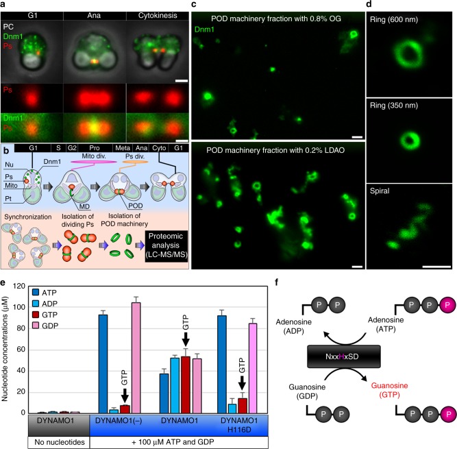
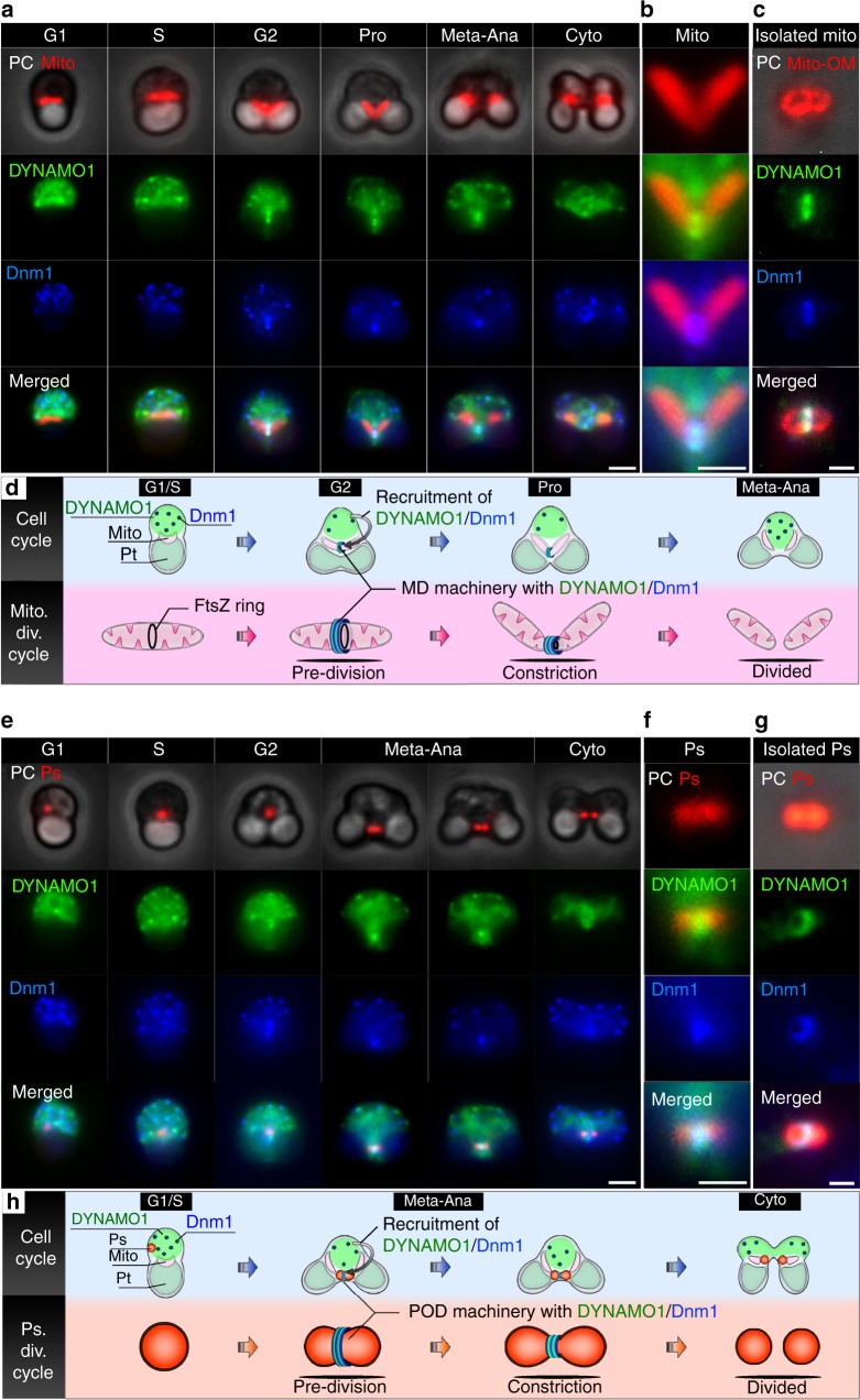
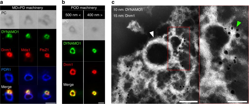
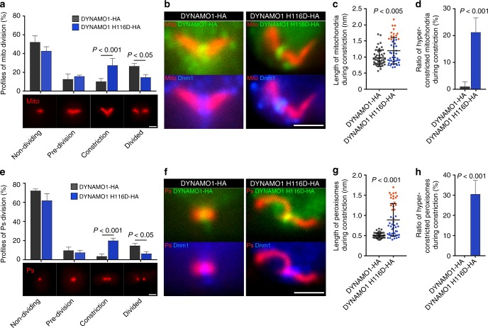
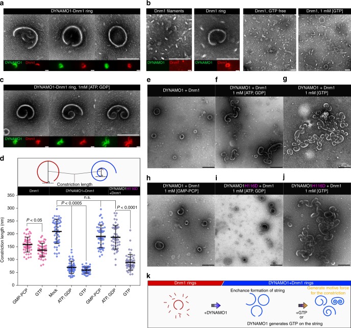
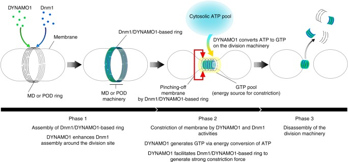
Mitochondria and peroxisomes proliferate by division. During division, a part of their membrane is pinched off by constriction of the ring-shaped mitochondrial division (MD) and peroxisome-dividing (POD) machinery. This constriction is mediated by a dynamin-like GTPase Dnm1 that requires a large amount of GTP as an energy source. Here, via proteomics of the isolated division machinery, we show that the 17-kDa nucleoside diphosphate kinase-like protein, dynamin-based ring motive-force organizer 1 (DYNAMO1), locally generates GTP in MD and POD machineries. DYNAMO1 is widely conserved among eukaryotes and colocalizes with Dnm1 on the division machineries. DYNAMO1 converts ATP to GTP, and disruption of its activity impairs mitochondrial and peroxisomal fissions. DYNAMO1 forms a ring-shaped complex with Dnm1 and increases the magnitude of the constricting force. Our results identify DYNAMO1 as an essential component of MD and POD machineries, suggesting that local GTP generation in Dnm1-based machinery regulates motive force for membrane severance.


Conflict of interest statement

The authors declare no competing interests.

Figures

Fig. 1
Fig. 1
Proteomic analysis of POD machinery and identification of DYNAMO1. a Phase contrast (PC) and immunofluorescence images of C. merolae cells during G1 phase, anaphase, and cytokinesis. Ps peroxisome (anti-catalase antibody), Dnm1 (anti-Dnm1 antibody). b Schematic representing isolation and proteomic analysis of POD machinery. Nu cell nucleus, Mito mitochondrion, Pt plastid, Mito div. mitochondrial division period, Ps div peroxisomal division period. c Upper panel shows the 0.8% OG-treated POD machinery fraction and the lower panel shows the 0.2% LDAO-treated POD machinery fraction. d Typical structures of isolated POD machinery stained with the anti-Dnm1 antibody. e LC–ESI–MS/MS analysis of the nucleoside diphosphate kinase activity of recombinant DYNAMO1. f Schematic representing a working model of nucleoside diphosphate kinase. Data in e are means ± s.d. (n = 3). Scale bars: 1 μm (a, upper panels); 500 nm (a, lower panels; c, d)
Fig. 2
Fig. 2
Dynamics of DYNAMO1 during the division periods of mitochondria and peroxisomes. a Phase contrast and immunofluorescence microscopy images of a mitochondrion (Mito, anti-EF-Tu antibody), DYNAMO1 (anti-DYNAMO1 antibody), and Dnm1 (anti-Dnm1 antibody) during the indicated cell cycle phases. Pro prophase, Meta–Ana metaphase–anaphase, Cyto cytokinesis. b Magnified image around the mitochondrial division site during prophase shown in a. c Localization of DYNAMO1 and Dnm1 on the isolated mitochondrial outer membrane [Mito-OM, anti-porin (POR) antibody]. d Schematic representing DYNAMO1 dynamics during the mitochondrial division period. e Phase contrast and immunofluorescence microscopy images of a peroxisome (Ps), DYNAMO1, and Dnm1 during the indicated cell cycle phases. f Magnified image around the peroxisomal division site during meta-anaphase shown in e. g Localization of DYNAMO1 and Dnm1 on an isolated dividing peroxisome. h Schematic representing DYNAMO1 dynamics during the peroxisomal division period. Scale bars: 1 μm (a, e); 500 nm (b, c, f, and g)
Fig. 3
Fig. 3
Localization of DYNAMO1 in MD and POD machineries. a Phase contrast and immunofluorescence microscopy images of DYNAMO1, Dnm1, Mda1 (anti-Mda1 antibody), FtsZ1 (anti-FtsZ1-antibody), and PDR1 (marker protein for PD machinery, anti-PDR1-antibody) on an isolated MD/PD machinery complex. b Phase contrast and immunofluorescence microscopy images of DYNAMO1 and Dnm1 on isolated POD machineries with diameters of >500 and <400 nm. c Whole-mount negative-staining immunoelectron microscopy images of DYNAMO1 (10 nm immunogold particles) and Dnm1 (15 nm immunogold particles) on isolated POD machinery. Right panel shows a magnified image of the red-boxed area in the left panel. The white arrowhead indicates a filamentous ring serving as a skeletal structure of POD machinery. The green arrowhead indicates a DB ring generating motive force for constriction. Scale bars: 1 μm (a); 500 nm (b); 200 nm (c); 50 nm (magnified image in c)
Fig. 4
Fig. 4
Disruption of DYNAMO1 functions and whole-mount negative-staining electron microscopy analysis of DYNAMO1-Dnm1 dynamics. a Profiles of mitochondrial division based on morphological changes during division. Mitochondria are labeled as in Fig. 2a. n = 3, at least 50 cells were counted in each experiment. b Localization of expressed DYNAMO1-HA and DYNAMO1 H116D-HA (Alexa 488-conjugated anti-HA antibody), Dnm1, and mitochondria in metaphase cells. c Length of mitochondria in cells expressing DYNAMO1-HA or DYNAMO1 H116D-HA. n = 50 in each experiment. Orange dots indicate hyper-constricted mitochondria as determined by quantification of their length that was elongated by more than 1.5 μm, which were positive for DYNAMO1 and Dnm1. d Ratio of hyper-constricted mitochondria during the constriction phase. e Profiles of peroxisomal division based on morphological changes during division. n = 3, at least 50 cells were counted in each experiment. f Localization of DYNAMO1-HA, DYNAMO1 H116D-HA, Dnm1, and peroxisomes in anaphase cells. g Lengths of peroxisomes in cells expressing DYNAMO1-HA or DYNAMO1 H116D-HA. n = 50 in each experiment. Orange dots indicate hyper-constricted peroxisomes as determined in c by quantification of their length that was elongated by more than 1 μm, which were positive for DYNAMO1 and Dnm1. h Ratio of hyper-constricted peroxisomes during the constriction phase. Scale bars: 1 μm. Data are means ± s.d. P; p-value (Mann–Whitney test)
Fig. 5
Fig. 5
Structure of DYNAMO1-Dnm1 string. a Typical structure of DYNAMO1-Dnm1 strings under GTP-free conditions. Immunofluorescence images of DYNAMO1 (green) and Dnm1 (red) are shown in the respective lower panels. b Typical structure of DYNAMO1-free Dnm1 filaments and strings under GTP-free condition (left, three panels) or in the presence of GTP (right panel). Immunofluorescence images are shown in the lower panels. c Typical structure of DYNAMO1-Dnm1 strings with addition of ATP and GDP. Immunofluorescence images are shown in the lower panels. d Schematic image representing measurement of constriction lengths of the strings. Constriction lengths of the DYNAMO1-free Dnm1, DYNAMO1-Dnm1, and DYNAMO1 H116D-Dnm1 strings under the various conditions (n = 50). ej Structural dynamics of DYNAMO1-Dnm1 or DYNAMO1 H116D-Dnm1 strings upon addition of ATP and GDP, GTP, or GMP-PCP. k Schematic image representing the dynamics of DYNAMO1-Dnm1 strings. Scale bars: 200 nm (ac); 500 nm (ej). Data are means ± s.d. n.s. not significant. P; p-value (unpaired t-test for Dnm1 and DYNAMO1 H116D + Dnm1, Kruskal–Wallis nonparametric ANOVA for DYNAMO1 + Dnm1)
Fig. 6
Fig. 6
A working model of DYNAMO1 functions during the division of mitochondrion and peroxisomes. Initially, DYNAMO1 is recruited to the division site of the mitochondrion, together with Dnm1 (Phase 1, prophase). DYNAMO1 is involved in Dnm1 assembly around the division site. Next, DYNAMO1 converts ATP to GTP on the ring of the MD machinery. A cytosolic ATP source is used for this reaction. Upon GTP generation, the MD machinery is constricted and GTPase Dnm1 pinches off mitochondrion. Moreover, the DYNAMO1-Dnm1 structure effectively generates a strong motive force for the constriction (Phase 2). MD machinery containing DYNAMO1 and Dnm1 is immediately disassembled (Phase 3). After mitochondrial division, DYNAMO1 functions in the POD machinery during meta-anaphase, in an analogous manner to that in mitochondrial division

References

    1. Danino D, Hinshaw JE. Dynamin family of mechanoenzymes. Curr. Opin. Cell Biol. 2001;13:454–460. doi: 10.1016/S0955-0674(00)00236-2. - DOI - PubMed
    1. Hinshaw JE, Schmid SL. Dynamin self-assembles into rings suggesting a mechanism for coated vesicle budding. Nature. 1995;374:690. doi: 10.1038/374190a0. - DOI - PubMed
    1. Bleazard W, et al. The dynamin-related GTPase Dnm1 regulates mitochondrial fission in yeast. Nat. Cell Biol. 1999;1:298–304. doi: 10.1038/13014. - DOI - PMC - PubMed
    1. Koch A, et al. Dynamin-like protein 1 is involved in peroxisomal fission. J. Biol. Chem. 2003;278:8597–8605. doi: 10.1074/jbc.M211761200. - DOI - PubMed
    1. Damke H, Baba T, Warnock DE, Schmid SL. Induction of mutant dynamin specifically blocks endocytic coated vesicle formation. J. Cell Biol. 1994;127:915–934. doi: 10.1083/jcb.127.4.915. - DOI - PMC - PubMed

Publication types